*Institute of Pharmacology, Catholic University Medical School, Rome, Italy.
†Institute of Pathology, Catholic University Medical School, Rome, Italy.
ASN Neuro. 2014 May 8;6(3):171-83. doi: 10.1042/AN20130045.
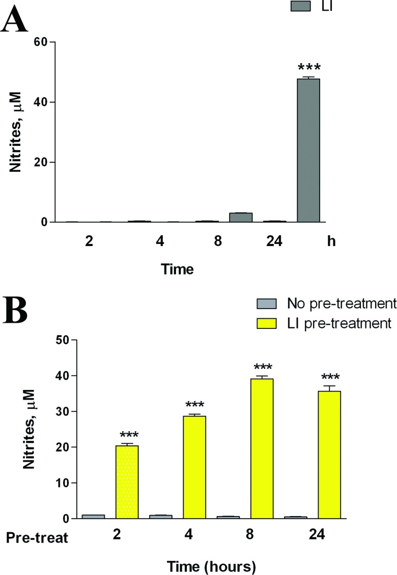
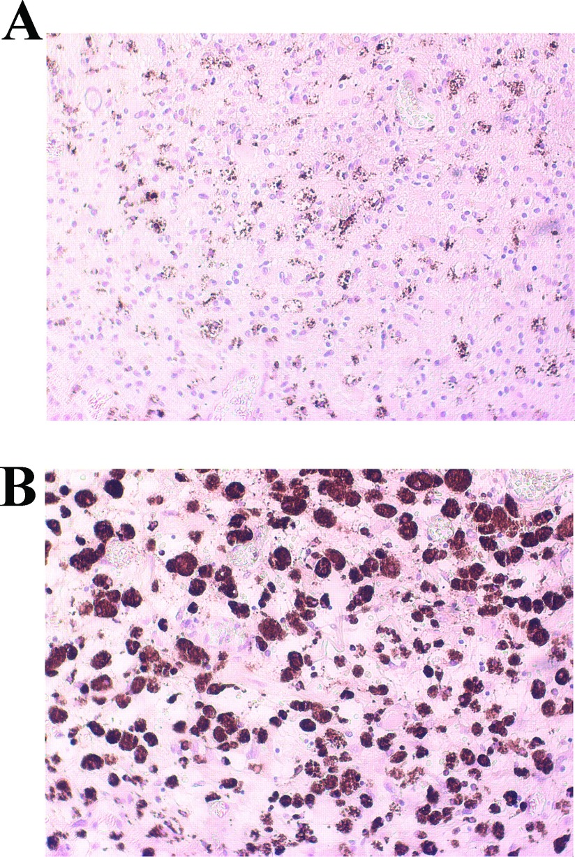
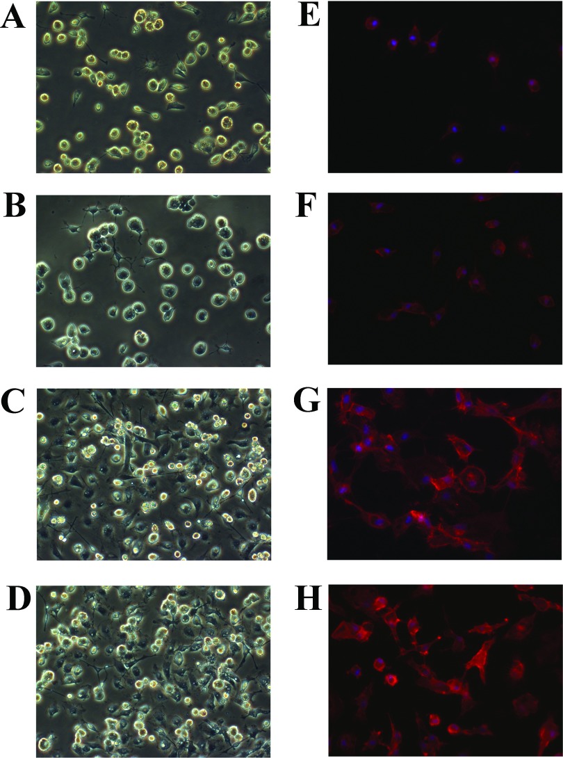
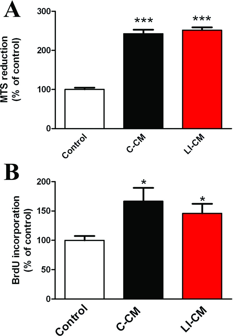
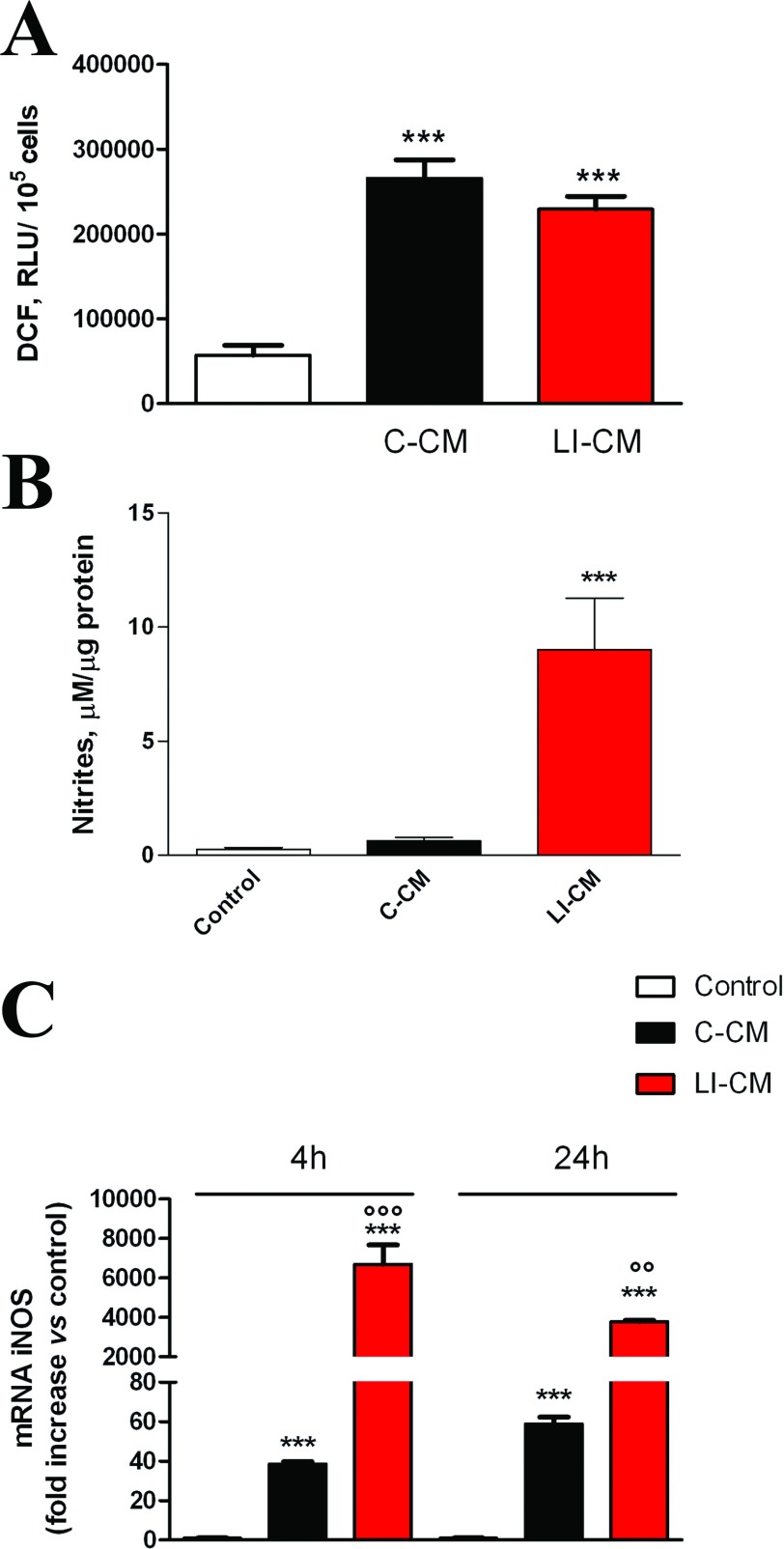
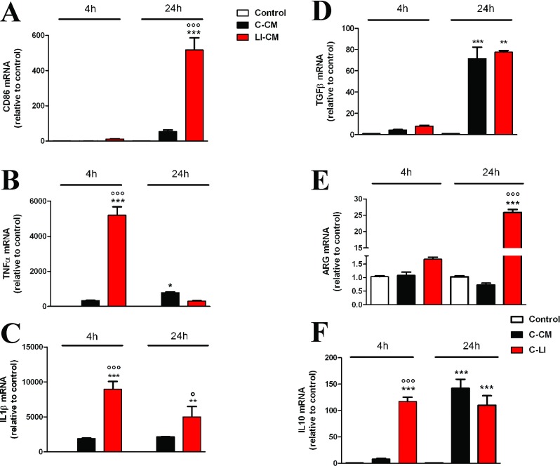
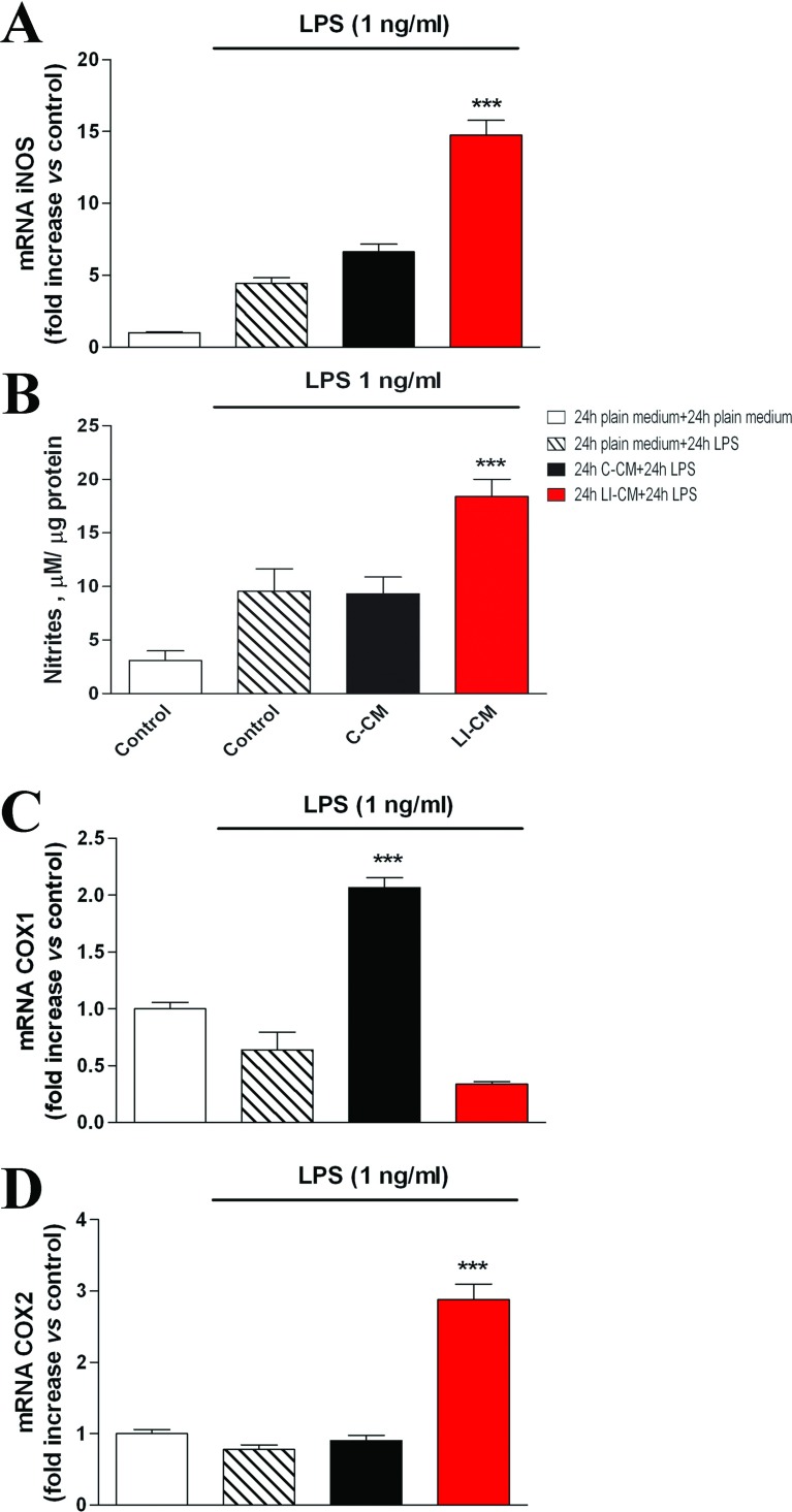
Malignant gliomas are primary brain tumors characterized by morphological and genetic complexities, as well as diffuse infiltration into normal brain parenchyma. Within gliomas, microglia/macrophages represent the largest tumor-infiltrating cell population, contributing by at least one-third to the total tumor mass. Bi-directional interactions between glioma cells and microglia may therefore play an important role on tumor growth and biology. In the present study, we have characterized the influence of glioma-soluble factors on microglial function, comparing the effects of media harvested under basal conditions with those of media obtained after inducing a pro-inflammatory activation state in glioma cells. We found that microglial cells undergo a different pattern of activation depending on the stimulus; in the presence of activated glioma-derived factors, i.e. a condition mimicking the late stage of pathology, microglia presents as a mixture of polarization phenotypes (M1 and M2a/b), with up-regulation of iNOS (inducible nitric oxide synthase), ARG (arginase) and IL (interleukine)-10. At variance, microglia exposed to basal glioma-derived factors, i.e. a condition resembling the early stage of pathology, shows a more specific pattern of activation, with increased M2b polarization status and up-regulation of IL-10 only. As far as viability and cell proliferation are concerned, both LI-CM [LPS (lipopolysaccharide)-IFNγ (interferon γ) conditioned media] and C-CM (control-conditioned media) induce similar effects on microglial morphology. Finally, in human glioma tissue obtained from surgical resection of patients with IV grade glioblastoma, we detected a significant amount of CD68 positive cells, which is a marker of macrophage/microglial phagocytic activity, suggesting that in vitro findings presented here might have a relevance in the human pathology as well.
恶性神经胶质瘤是原发性脑肿瘤,其特征在于形态和遗传复杂性,以及弥漫性浸润正常脑实质。在神经胶质瘤中,小胶质细胞/巨噬细胞代表最大的肿瘤浸润细胞群体,至少占肿瘤总质量的三分之一。因此,神经胶质瘤细胞与小胶质细胞之间的双向相互作用可能对肿瘤生长和生物学特性起着重要作用。在本研究中,我们对神经胶质瘤可溶性因子对小胶质细胞功能的影响进行了特征描述,将基础条件下收集的培养基的影响与诱导神经胶质瘤细胞产生促炎激活状态后获得的培养基的影响进行了比较。我们发现,小胶质细胞根据刺激的不同会呈现出不同的激活模式;在存在激活的神经胶质瘤衍生因子的情况下,即模拟病理晚期的条件下,小胶质细胞呈现出极化表型(M1 和 M2a/b)的混合物,诱导型一氧化氮合酶(iNOS)、精氨酸酶(ARG)和白细胞介素(IL)-10 的表达上调。相反,暴露于基础神经胶质瘤衍生因子的小胶质细胞,即类似于病理早期的条件,显示出更特异的激活模式,M2b 极化状态增加,仅上调 IL-10。就活力和细胞增殖而言,LI-CM[脂多糖(LPS)-干扰素γ(IFNγ)条件培养基]和 C-CM(对照条件培养基)对小胶质细胞形态均有相似的诱导作用。最后,在从患有 IV 级神经胶质瘤的患者的手术切除的人神经胶质瘤组织中,我们检测到大量 CD68 阳性细胞,这是巨噬细胞/小胶质细胞吞噬活性的标志物,表明此处呈现的体外发现可能与人的病理学也有关。