Clements P R, Brooks D A, McCourt P A, Hopwood J J
Department of Chemical Pathology, Adelaide Children's Hospital, Australia.
Biochem J. 1989 Apr 1;259(1):199-208. doi: 10.1042/bj2590199.
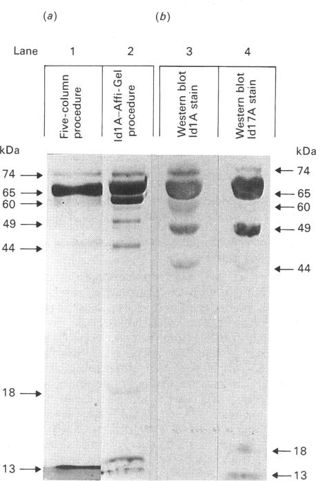
alpha-L-Iduronidase from human liver was purified by a three-step five-column procedure and by immunoaffinity chromatography with a monoclonal antibody raised against purified enzyme. Seven bands identified by staining with Coomassie Blue had molecular masses of 74, 65, 60, 49, 44, 18 and 13 kDa and were present in both preparations of the liver enzyme. However, relative to the immunopurification procedure, alpha-L-iduronidase purified by the five-column procedure was considerably enriched in the 65 kDa polypeptide band. The seven bands were identified by Western-blot analysis with two different monoclonal antibodies raised against alpha-L-iduronidase. The chromatographic behaviour of alpha-L-iduronidase on the antibody column was dependent upon the quantity of enzyme loaded. Above a particular load concentration a single peak of enzyme activity was eluted, whereas at load concentrations below the critical value alpha-L-iduronidase was eluted in two peaks of activity, designated form I (eluted first) and form II (eluted second). The following properties of the two forms of alpha-L-iduronidase were determined. (1) The two forms from liver were composed of different proportions of the same seven polypeptides. (2) When individually rechromatographed on the antibody column, each form from liver shifted to a more retarded elution position but essentially retained its chromatographic behaviour relative to the other form. (3) Forms I and II of liver alpha-L-iduronidase showed no difference in their activities towards disaccharide substrates derived from two glycosaminoglycan sources, heparan sulphate and dermatan sulphate. (4) The native molecular size of forms I and II of liver alpha-L-iduronidase was 65 kDa as determined by gel-permeation chromatography. (5) Immunoaffinity chromatography of extracts of human lung and kidney resulted in the separation of alpha-L-iduronidase into two forms, each with different proportions of the seven common polypeptide species. (6) Lung forms I and II were taken up readily into cultured skin fibroblasts taken from a patient with alpha-L-iduronidase deficiency. Liver forms I and II were not taken up to any significant extent. Lung form II gave intracellular contents of alpha-L-iduronidase that were more than double those of normal control fibroblasts, whereas lung form I gave contents approximately equal to normal control values. We propose that all seven polypeptides are derived from a single alpha-L-iduronidase gene product, and that different proportions of these polypeptides can function as a single alpha-L-iduronidase entity.(ABSTRACT TRUNCATED AT 400 WORDS)
人肝脏中的α-L-艾杜糖醛酸酶通过三步五柱法以及使用针对纯化酶产生的单克隆抗体进行免疫亲和层析进行纯化。用考马斯亮蓝染色鉴定出的七条带的分子量分别为74、65、60、49、44、18和13 kDa,且在肝脏酶的两种制剂中均存在。然而,相对于免疫纯化方法,通过五柱法纯化的α-L-艾杜糖醛酸酶在65 kDa多肽带中显著富集。用两种针对α-L-艾杜糖醛酸酶产生的不同单克隆抗体通过蛋白质印迹分析鉴定出这七条带。α-L-艾杜糖醛酸酶在抗体柱上的色谱行为取决于加载的酶量。在特定加载浓度以上,洗脱的是单一的酶活性峰,而在低于临界值的加载浓度下,α-L-艾杜糖醛酸酶以两个活性峰洗脱,分别命名为I型(先洗脱)和II型(后洗脱)。测定了α-L-艾杜糖醛酸酶两种形式的以下特性。(1)来自肝脏的两种形式由相同的七种多肽以不同比例组成。(2)当在抗体柱上单独重新层析时,来自肝脏的每种形式都迁移到更滞后的洗脱位置,但相对于另一种形式基本上保留其色谱行为。(3)肝脏α-L-艾杜糖醛酸酶的I型和II型对源自两种糖胺聚糖来源(硫酸乙酰肝素和硫酸皮肤素)的二糖底物的活性没有差异。(4)通过凝胶渗透色谱法测定,肝脏α-L-艾杜糖醛酸酶I型和II型的天然分子大小为65 kDa。(5)人肺和肾提取物的免疫亲和层析导致α-L-艾杜糖醛酸酶分离为两种形式,每种形式中七种常见多肽种类的比例不同。(6)肺I型和II型很容易被取自α-L-艾杜糖醛酸酶缺乏症患者的培养皮肤成纤维细胞摄取。肝脏I型和II型没有被显著摄取。肺II型产生的α-L-艾杜糖醛酸酶细胞内含量是正常对照成纤维细胞的两倍多,而肺I型产生的含量约等于正常对照值。我们提出所有七种多肽均源自单一的α-L-艾杜糖醛酸酶基因产物,并且这些多肽的不同比例可以作为单一的α-L-艾杜糖醛酸酶实体发挥作用。(摘要截短至400字)