Chen Huisong, Trilok Goolab, Wang Fei, Qi Xiaolong, Xiao Junjie, Yang Changqing
Division of Gastroenterology & Hepatology, Digestive Disease Institute, Tongji Hospital, Tongji University School of Medicine, Shanghai, China.
Indian J Med Res. 2014 Feb;139(2):260-6.
BACKGROUND & OBJECTIVES: Discrepancies exist in the reported prevalence of portal vein thrombosis (PVT), and its clinical characteristics and sites of occurrence need to be elucidated. The risk factors for PVT are also poorly understood. This single centre study was undertaken to determine the clinical characteristics, sites of occurrence, and risk factors associated with PVT in patients with liver cirrhosis.
Hospitalized cirrhotic patients (N = 162) were segregated into the PVT and non-PVT groups. Indices possibly associated with PVT were measured and PVT was detected by both Doppler ultrasonography and computed tomography portal angiography. The portal vein diameter and flow velocity and splenic thickness were measured by ultrasonography.
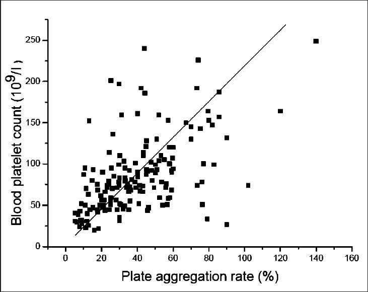
PVT was found in 40 patients (24.7%); in 34 PVT patients (85%), the liver cirrhosis resulted from hepatitis B virus infections. Most (90%) patients were Child-Pugh classes B and C, with similar distribution between the groups. PVT was seen in 20 patients in the portal and superior mesenteric veins; ascites, abdominal pain, gastrointestinal bleeding, and jaundice were common findings in PVT patients. Haemoglobin levels and blood platelet counts (BPCs) were significantly lower and splenic thickness was greater in PVT than in non-PVT patients (P<0.01). There was a significant positive correlation between BPCs and platelet aggregation rates (R = 0.533, P<0.01).
INTERPRETATION & CONCLUSIONS: The occurrence of PVT was 24.7 per cent, primarily in post-hepatitis B liver cirrhosis patients. PVT occurred mainly in the portal vein trunk and superior mesenteric vein. Different PVT sites may account for the differing clinical presentations. The lower levels of haemoglobin and BPCs as well as splenic thickening were associated with PVT. Splenic thickening may be a risk factor for PVT.
门静脉血栓形成(PVT)的报道患病率存在差异,其临床特征及发生部位有待阐明。PVT的危险因素也尚未完全明确。本单中心研究旨在确定肝硬化患者中PVT的临床特征、发生部位及相关危险因素。
将162例住院肝硬化患者分为PVT组和非PVT组。测量可能与PVT相关的指标,并通过多普勒超声和CT门静脉血管造影检测PVT。用超声测量门静脉直径、血流速度和脾厚度。
40例患者(24.7%)发现有PVT;34例PVT患者(85%)的肝硬化由乙型肝炎病毒感染引起。大多数(90%)患者为Child-Pugh B级和C级,两组间分布相似。20例患者门静脉和肠系膜上静脉出现PVT;腹水、腹痛、胃肠道出血和黄疸是PVT患者的常见表现。PVT患者的血红蛋白水平和血小板计数(BPC)显著低于非PVT患者,脾厚度更大(P<0.01)。BPC与血小板聚集率之间存在显著正相关(R = 0.533,P<0.01)。
PVT的发生率为24.7%,主要见于乙型肝炎后肝硬化患者。PVT主要发生在门静脉主干和肠系膜上静脉。不同的PVT部位可能导致不同的临床表现。血红蛋白和BPC水平降低以及脾增厚与PVT有关。脾增厚可能是PVT的一个危险因素。