Ziółkowska Natalia, Lewczuk Bogdan, Petryński Wojciech, Palkowska Katarzyna, Prusik Magdalena, Targońska Krystyna, Giżejewski Zygmunt, Przybylska-Gornowicz Barbara
Department of Histology and Embryology, Faculty of Veterinary Medicine, University of Warmia and Mazury, Olsztyn, Poland.
2Institute of Animal Reproduction and Food Research of Polish Academy of Sciences, Olsztyn, Poland.
PLoS One. 2014 Apr 11;9(4):e94590. doi: 10.1371/journal.pone.0094590. eCollection 2014.
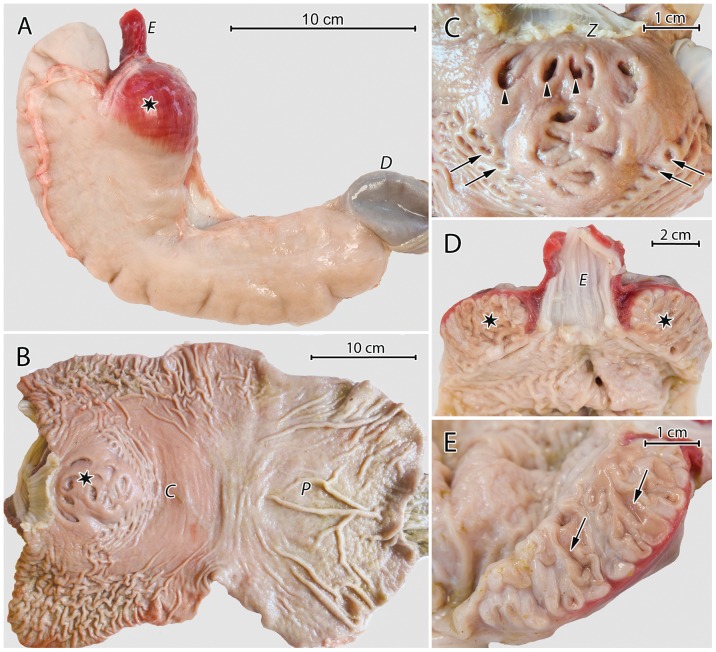
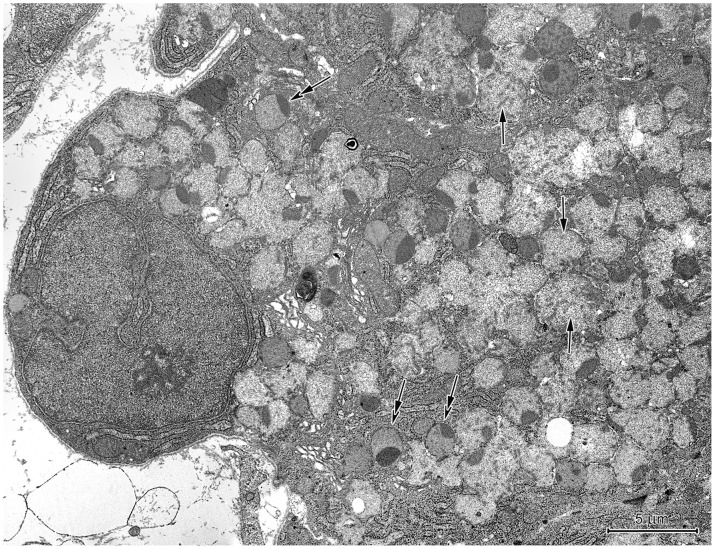
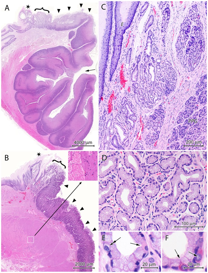
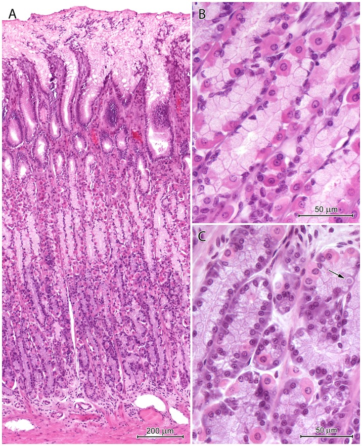
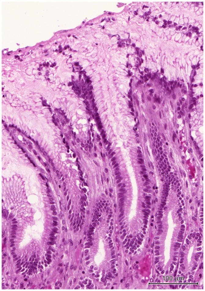
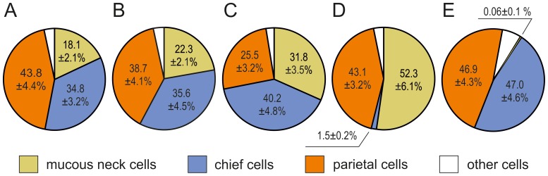
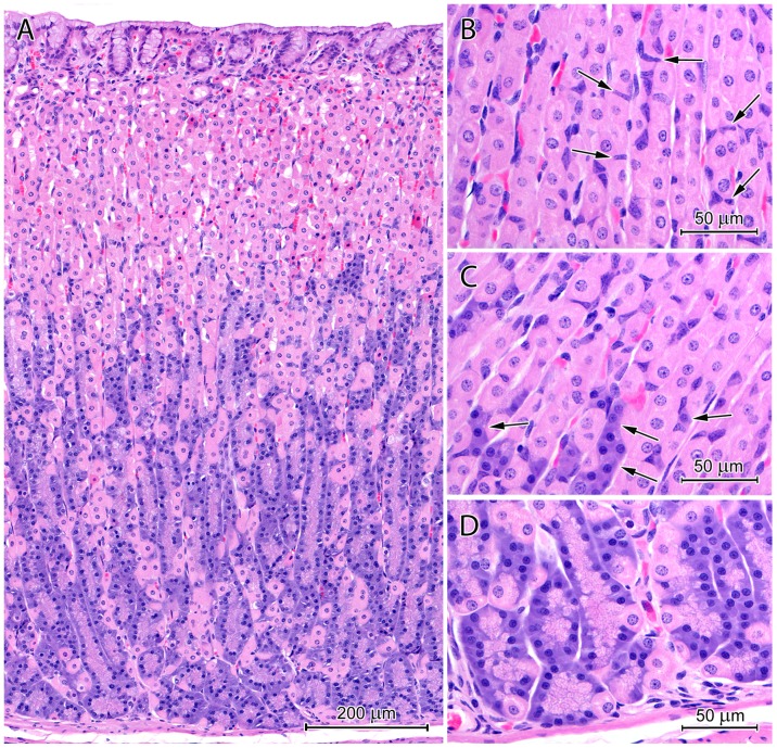
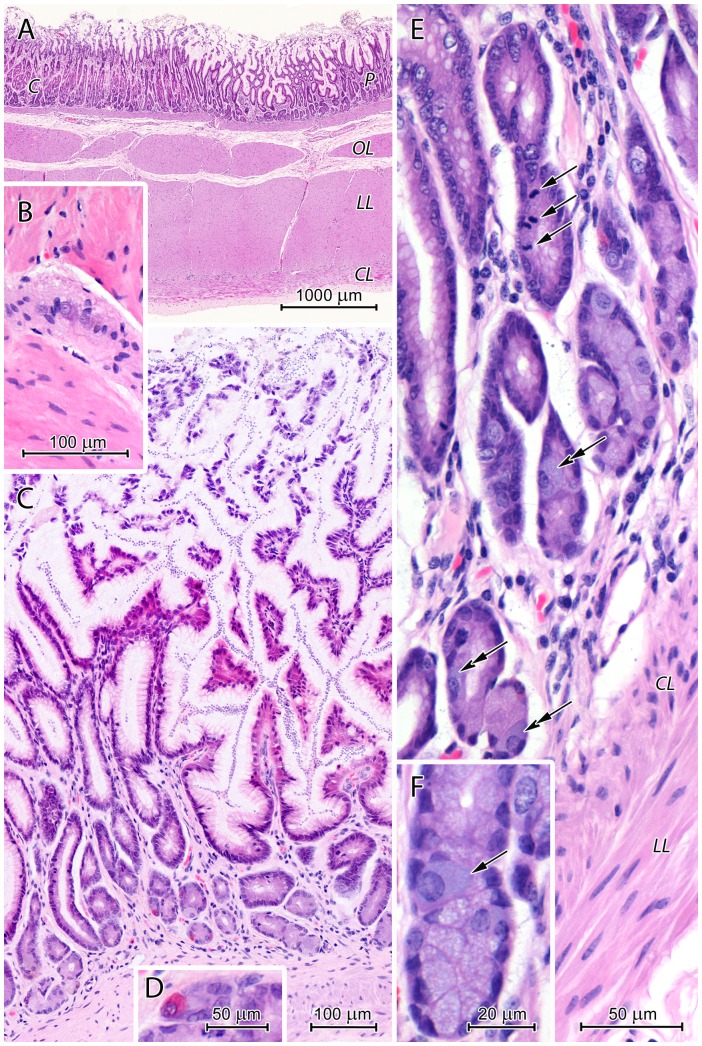
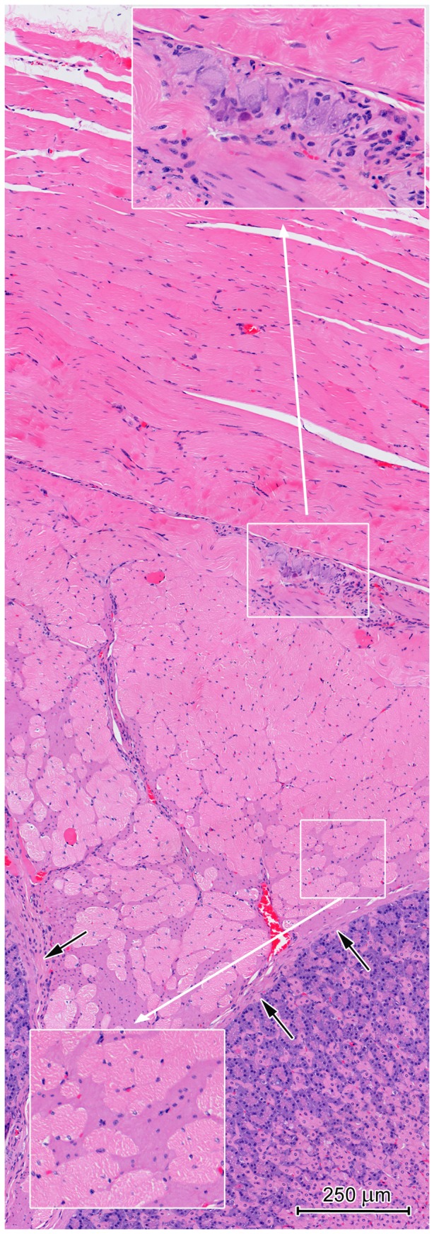
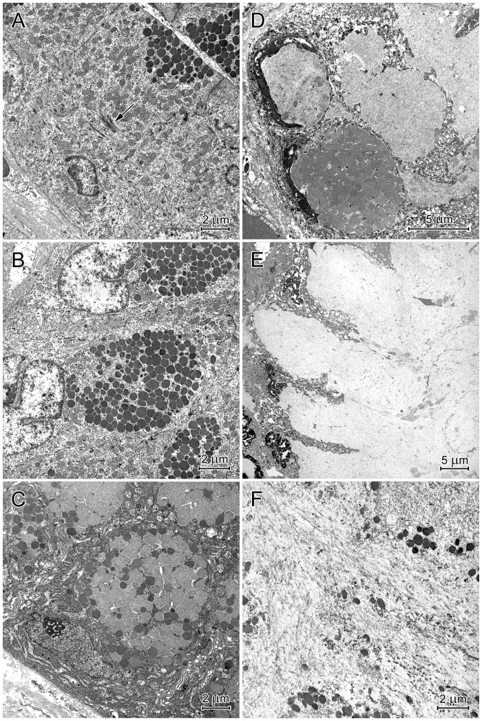
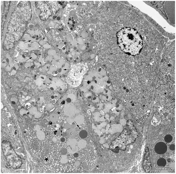
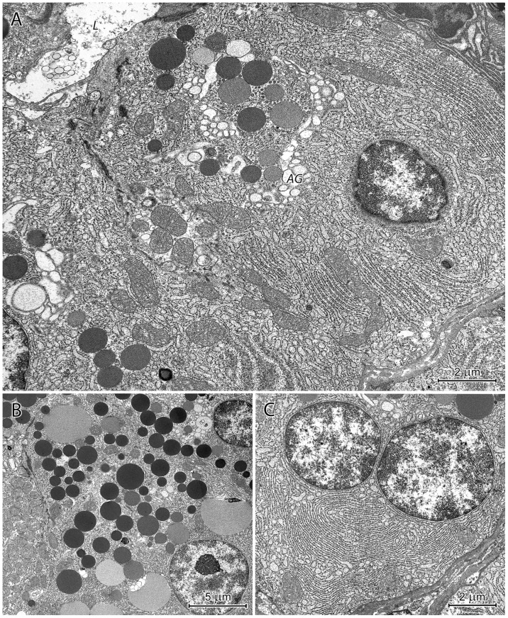
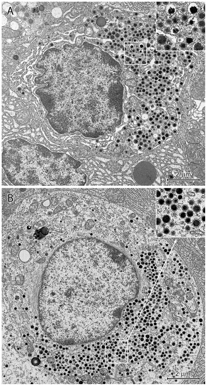
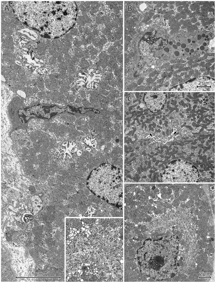
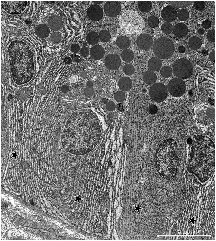
Anatomical, histological, and ultrastructural studies of the European beaver stomach revealed several unique morphological features. The prominent attribute of its gross morphology was the cardiogastric gland (CGG), located near the oesophageal entrance. Light microscopy showed that the CGG was formed by invaginations of the mucosa into the submucosa, which contained densely packed proper gastric glands comprised primarily of parietal and chief cells. Mucous neck cells represented <0.1% of cells in the CGG gastric glands and 22-32% of cells in the proper gastric glands of the mucosa lining the stomach lumen. These data suggest that chief cells in the CGG develop from undifferentiated cells that migrate through the gastric gland neck rather than from mucous neck cells. Classical chief cell formation (i.e., arising from mucous neck cells) occurred in the mucosa lining the stomach lumen, however. The muscularis around the CGG consisted primarily of skeletal muscle tissue. The cardiac region was rudimentary while the fundus/corpus and pyloric regions were equally developed. Another unusual feature of the beaver stomach was the presence of specific mucus with a thickness up to 950 µm (in frozen, unfixed sections) that coated the mucosa. Our observations suggest that the formation of this mucus is complex and includes the secretory granule accumulation in the cytoplasm of pit cells, the granule aggregation inside cells, and the incorporation of degenerating cells into the mucus.
对欧洲河狸胃的解剖学、组织学和超微结构研究揭示了几个独特的形态学特征。其大体形态的突出特征是位于食管入口附近的心胃腺(CGG)。光学显微镜显示,心胃腺由黏膜向黏膜下层内陷形成,其中含有密集排列的固有胃腺,主要由壁细胞和主细胞组成。黏液颈细胞在心胃腺胃腺细胞中占比不到0.1%,在胃腔内衬黏膜的固有胃腺细胞中占比22 - 32%。这些数据表明,心胃腺中的主细胞由未分化细胞发育而来,这些未分化细胞通过胃腺颈部迁移,而非由黏液颈细胞发育而来。然而,胃腔内衬黏膜中发生了典型的主细胞形成过程(即由黏液颈细胞产生)。心胃腺周围的肌层主要由骨骼肌组织组成。贲门区发育不全,而胃底/胃体和幽门区发育程度相当。河狸胃的另一个不寻常特征是存在一种特定的黏液,其厚度可达950微米(在冷冻、未固定切片中),覆盖在黏膜上。我们的观察结果表明,这种黏液的形成过程很复杂,包括在凹细胞的细胞质中积累分泌颗粒、细胞内颗粒聚集以及将退化细胞纳入黏液中。