Castonguay Alexandre, Thomas Sébastien, Lesage Frédéric, Casanova Christian
École d'optométrie, Université de Montréal, CP 6128 succursale centre-ville, Montréal, Quebec, Canada.
École Polytechnique de Montréal, CP 6079 succursale centre-ville, Montréal, Quebec, Canada.
PLoS One. 2014 Apr 11;9(4):e94633. doi: 10.1371/journal.pone.0094633. eCollection 2014.
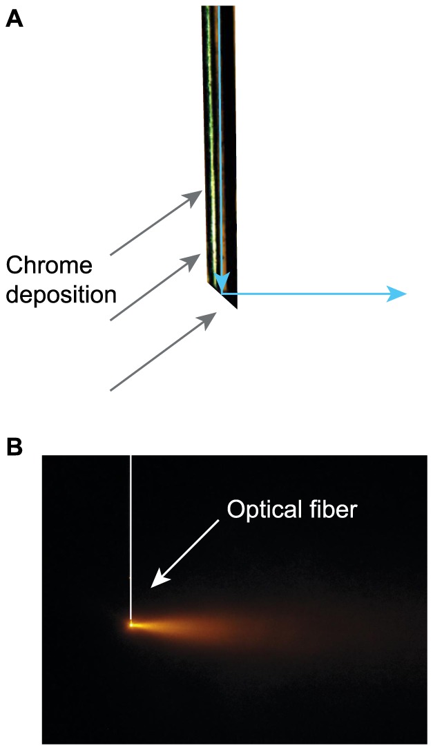
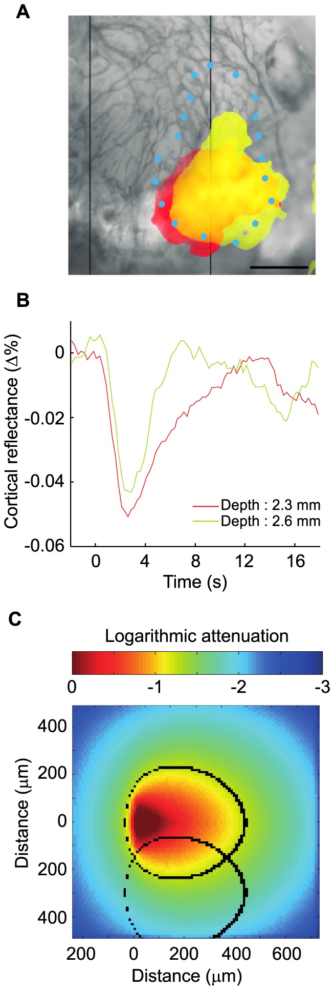
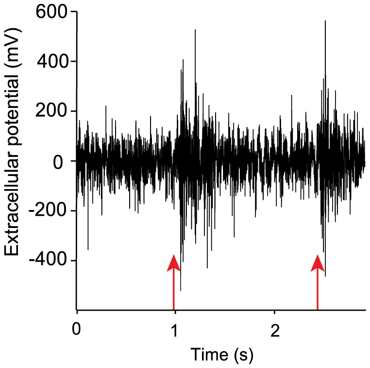
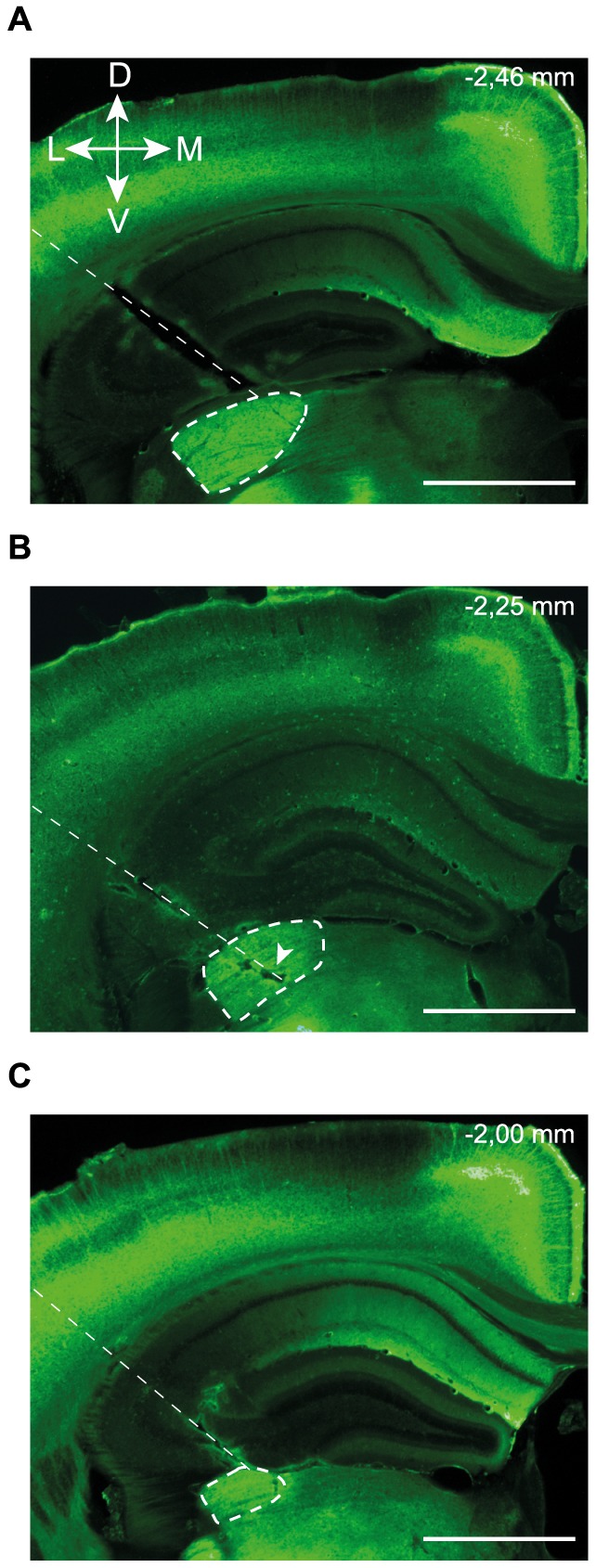
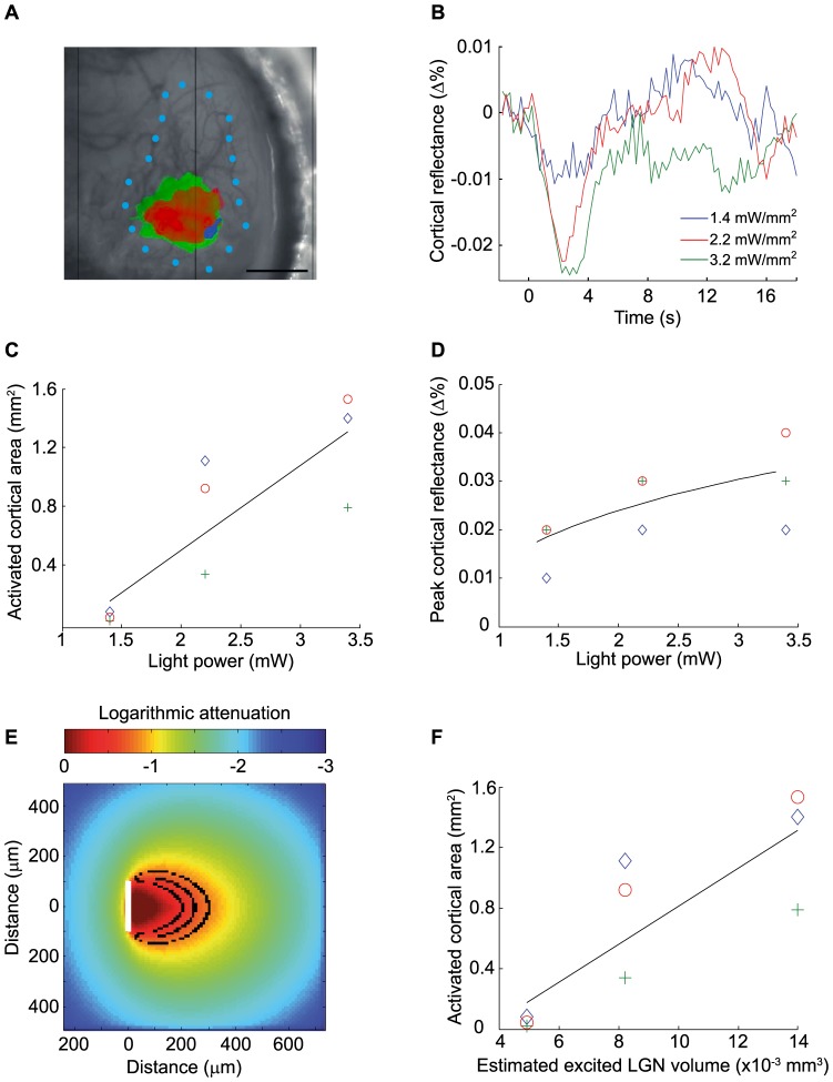
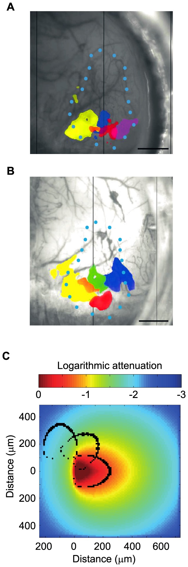
Optogenetics allows the control of cellular activity using focused delivery of light pulses. In neuroscience, optogenetic protocols have been shown to efficiently inhibit or stimulate neuronal activity with a high temporal resolution. Among the technical challenges associated with the use of optogenetics, one is the ability to target a spatially specific population of neurons in a given brain structure. To address this issue, we developed a side-illuminating optical fiber capable of delivering light to specific sites in a target nucleus with added flexibility through rotation and translation of the fiber and by varying the output light power. The designed optical fiber was tested in vivo in visual structures of ChR2-expressing transgenic mice. To assess the spatial extent of neuronal activity modulation, we took advantage of the hallmark of the visual system: its retinotopic organization. Indeed, the relative position of ganglion cells in the retina is transposed in the cellular topography of both the dorsal lateral geniculate nucleus (LGN) in the thalamus and the primary visual cortex (V1). The optical fiber was inserted in the LGN and by rotating it with a motor, it was possible to sequentially activate different neuronal populations within this structure. The activation of V1 neurons by LGN projections was recorded using intrinsic optical imaging. Increasing light intensity (from 1.4 to 8.9 mW/mm²) led to increasing activation surfaces in V1. Optogenetic stimulation of the LGN at different translational and rotational positions was associated with different activation maps in V1. The position and/or orientation of the fiber inevitably varied across experiments, thus limiting the capacity to pool data. With the optogenetic design presented here, we demonstrate for the first time a transitory and spatially-concise activation of a deep neuronal structure. The optogenetic design presented here thus opens a promising avenue for studying the function of deep brain structures.
光遗传学可通过聚焦递送光脉冲来控制细胞活动。在神经科学领域,光遗传学方案已被证明能够以高时间分辨率有效抑制或刺激神经元活动。在与光遗传学应用相关的技术挑战中,一个挑战是能够在给定脑结构中靶向特定空间的神经元群体。为了解决这个问题,我们开发了一种侧照式光纤,它能够通过光纤的旋转和平移以及改变输出光功率,将光灵活地递送至目标核中的特定部位。所设计的光纤在表达ChR2的转基因小鼠的视觉结构中进行了体内测试。为了评估神经元活动调节的空间范围,我们利用了视觉系统的特征:其视网膜拓扑组织。实际上,视网膜中神经节细胞的相对位置在丘脑背侧外侧膝状体核(LGN)和初级视觉皮层(V1)的细胞拓扑结构中得以转换。将光纤插入LGN,并通过电机旋转光纤,可以依次激活该结构内不同的神经元群体。使用内在光学成像记录LGN投射对V1神经元的激活情况。增加光强度(从1.4到8.9毫瓦/平方毫米)会导致V1中激活表面增加。在不同平移和旋转位置对LGN进行光遗传学刺激与V1中不同的激活图谱相关。在不同实验中,光纤的位置和/或方向不可避免地会发生变化,从而限制了汇总数据的能力。通过本文提出的光遗传学设计,我们首次证明了对深部神经元结构的短暂且空间精确的激活。因此,本文提出的光遗传学设计为研究深部脑结构的功能开辟了一条有前景的途径。