Department of Comparative Biomedicine and Food Science, University of Padova Legnaro, Italy.
Department of Psychology, Catholic University Milan, Italy.
Front Syst Neurosci. 2014 Mar 28;8:42. doi: 10.3389/fnsys.2014.00042. eCollection 2014.
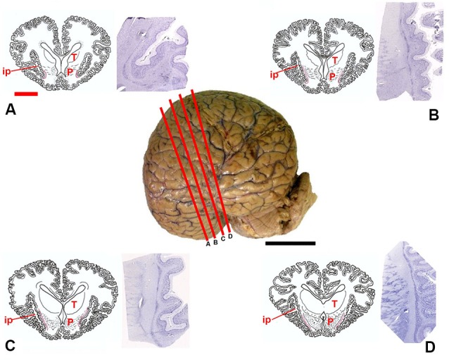
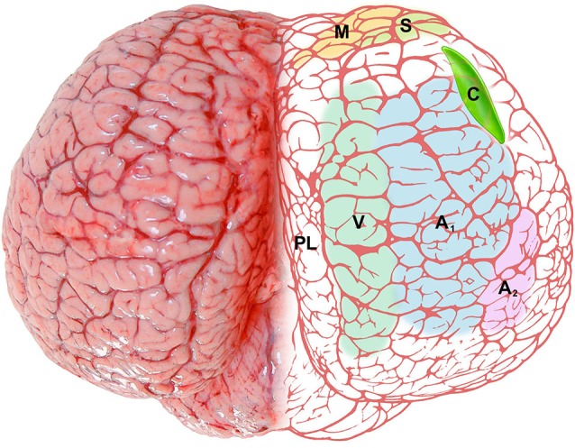
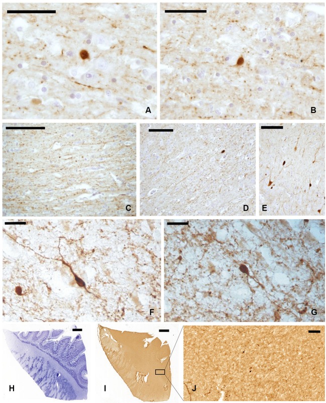
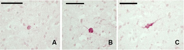
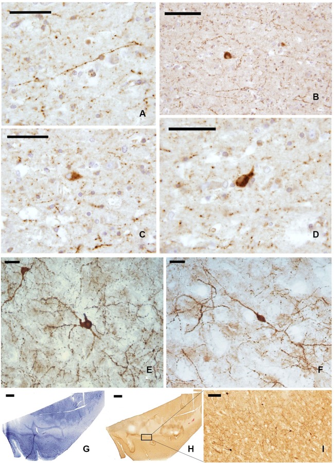
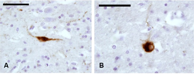
The mammalian claustrum is involved in processing sensory information from the environment. The claustrum is reciprocally connected to the visual cortex and these projections, at least in carnivores, display a clear retinotopic distribution. The visual cortex of dolphins occupies a position strikingly different from that of land mammals. Whether the reshaping of the functional areas of the cortex of cetaceans involves also modifications of the claustral projections remains hitherto unanswered. The present topographic and immunohistochemical study is based on the brains of eight bottlenose dolphins and a wide array of antisera against: calcium-binding proteins (CBPs) parvalbumin (PV), calretinin (CR), and calbindin (CB); somatostatin (SOM); neuropeptide Y (NPY); and the potential claustral marker Gng2. Our observations confirmed the general topography of the mammalian claustrum also in the bottlenose dolphin, although (a) the reduction of the piriform lobe modifies the ventral relationships of the claustrum with the cortex, and (b) the rotation of the telencephalon along the transverse axis, accompanied by the reduction of the antero-posterior length of the brain, apparently moves the claustrum more rostrally. We observed a strong presence of CR-immunoreactive (-ir) neurons and fibers, a diffuse but weak expression of CB-ir elements and virtually no PV immunostaining. This latter finding agrees with studies that report that PV-ir elements are rare in the visual cortex of the same species. NPY- and somatostatin-containing neurons were evident, while the potential claustral markers Gng2 was not identified in the sections, but no explanation for its absence is currently available. Although no data are available on the projections to and from the claustrum in cetaceans, our results suggest that its neurochemical organization is compatible with the presence of noteworthy cortical inputs and outputs and a persistent role in the general processing of the relative information.
哺乳动物的屏状核参与处理来自环境的感觉信息。屏状核与视皮层相互连接,这些投射至少在食肉动物中表现出明显的视网膜分布。海豚的视皮层占据的位置与陆地哺乳动物明显不同。鲸类动物皮层功能区的重塑是否还涉及屏状核投射的改变,目前仍未得到解答。本研究基于 8 只宽吻海豚的大脑进行,使用了多种针对:钙结合蛋白(CBPs)- 钙结合蛋白 28k(PV)、钙调蛋白(CR)和钙结合蛋白 2(CB);生长抑素(SOM);神经肽 Y(NPY);和潜在的屏状核标记 Gng2 的抗体。我们的观察结果证实了在宽吻海豚中也存在哺乳动物屏状核的一般拓扑结构,尽管(a)梨状叶的减小改变了屏状核与皮层的腹侧关系,(b)沿着横向轴的大脑旋转,伴随着大脑前后长度的减小,显然使屏状核向更前端移动。我们观察到 CR-免疫反应性(-ir)神经元和纤维的强烈存在,CB-ir 元素的弥散但微弱表达和实际上没有 PV 免疫染色。这一发现与报告相同物种的视皮层中 PV-ir 元素罕见的研究结果一致。存在 NPY 和生长抑素神经元,而潜在的屏状核标记 Gng2 在切片中未被识别,但目前尚无其不存在的解释。尽管没有关于鲸类动物屏状核的投射的信息,但我们的结果表明,其神经化学组织与存在显著的皮质输入和输出以及在相对信息的一般处理中持续发挥作用是兼容的。