Department of Anatomy, Charles University in Prague, 2nd Faculty of Medicine Prague, Czech Republic ; Department of Anatomy, Charles University in Prague, 1st Faculty of Medicine Prague, Czech Republic ; Department of Developmental Epileptology, Institute of Physiology, Academy of Sciences of the Czech Republic Prague, Czech Republic.
Department of Anatomy, Charles University in Prague, 2nd Faculty of Medicine Prague, Czech Republic.
Front Neuroanat. 2015 Jan 20;8:160. doi: 10.3389/fnana.2014.00160. eCollection 2014.
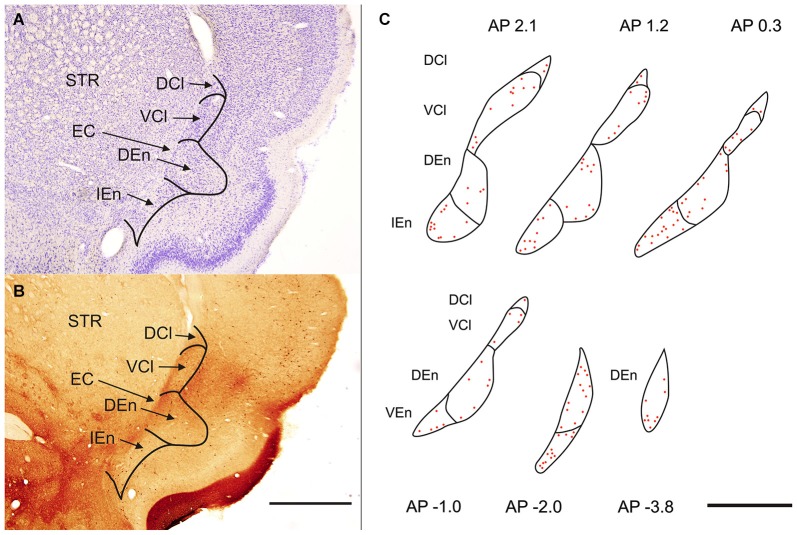
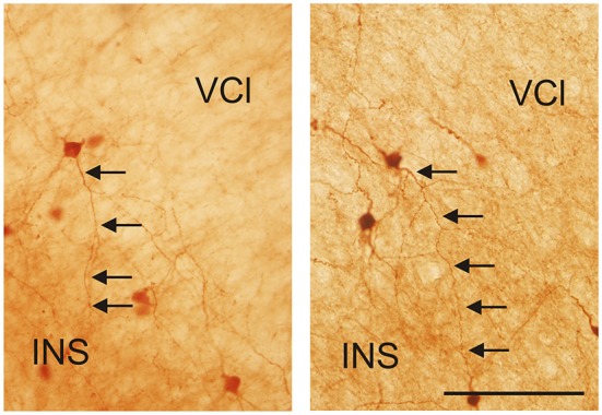
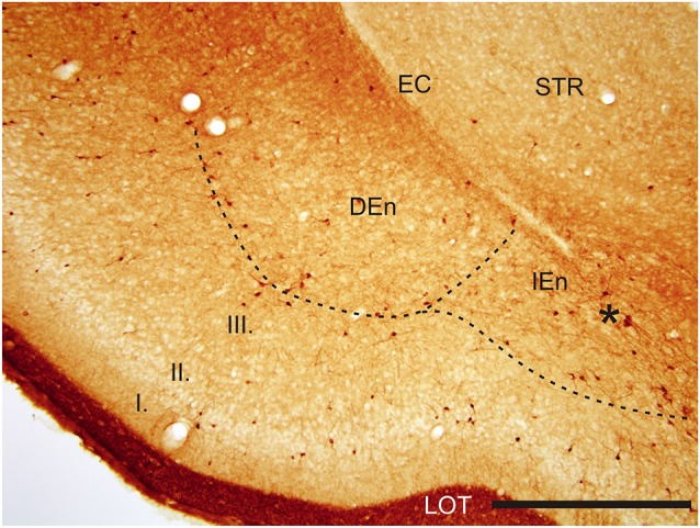
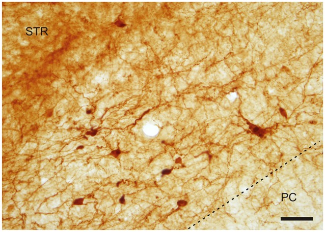
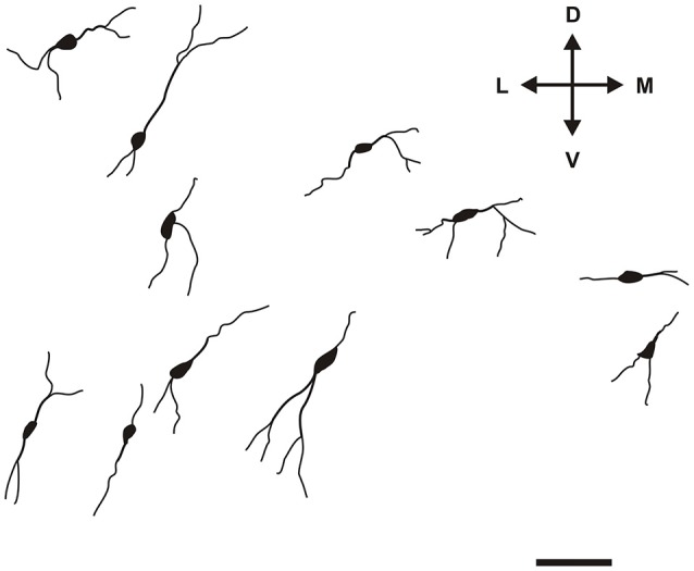
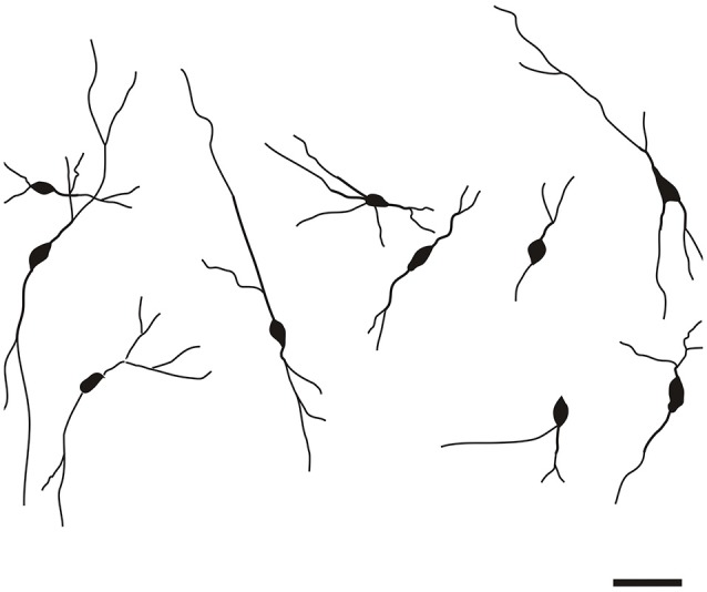
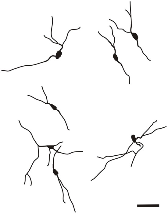
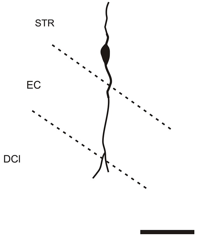
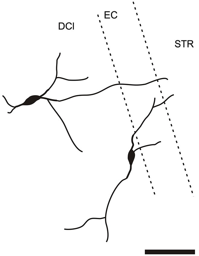
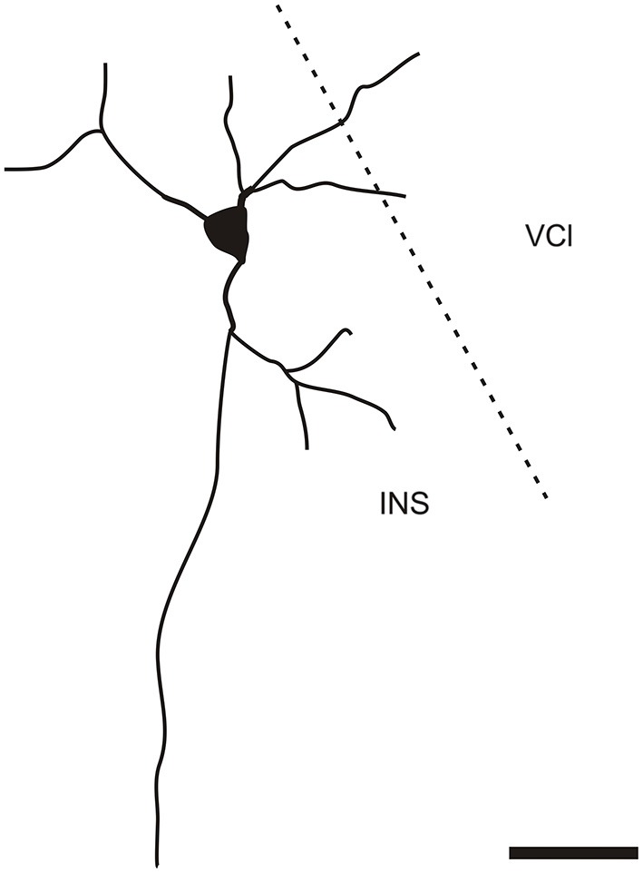
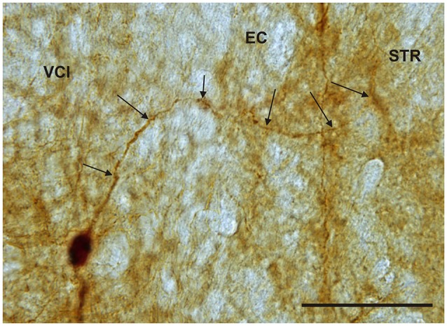
The claustrum is a telencephalic structure which consists of dorsal segment adjoining the insular cortex and a ventral segment termed also endopiriform nucleus (END). The dorsal segment (claustrum) is divided into a dorsal and ventral zone, while the END is parcellated into dorsal, ventral and intermediate END. The claustrum and the END consist of glutamatergic projection neurons and GABAergic local interneurons coexpressing calcium binding proteins. Among neurons expressing calcium binding proteins the calretinin (CR)-immunoreactive interneurons exert specific functions in neuronal circuits, including disinhibition of excitatory neurons. Previous anatomical data indicate extensive and reciprocally organized claustral projections with cerebral cortex. We asked if the distribution of cells immunoreactive for CR delineates anatomical or functional subdivisions in the claustrum and in the END. Both segments of the claustrum and all subdivisions of the END contained CR immunoreactive neurons with varying distribution. The ventral zone of the claustrum exhibited weak labeling with isolated cell bodies and thin fibers and is devoid of immunoreactive puncta. Within the medial margin of the intermediate END we noted a group of strongly positive neurons. Cells immunoreactive for CR in all subdivisions of the claustrum and END were bipolar, multipolar and oval with smooth, beaded aspiny dendrites. Small number of CR-immunoreactive neurons displayed thin dendrites which enter to adjoining structures. Penetration of dendrites was reciprocal. These results show an inhomogenity over the claustrum and the END in distribution and types of CR immunoreactive neurons. The distribution of the CR-immunoreactive neurons respects the anatomical but not functional zones of the claustral complex.
屏状核是端脑结构,由毗邻脑岛的背侧节段和称为内嗅核(END)的腹侧节段组成。背侧节段(屏状核)分为背侧和腹侧区,而 END 则分为背侧、腹侧和中间 END。屏状核和 END 由谷氨酸能投射神经元和 GABA 能局部中间神经元组成,这些神经元共表达钙结合蛋白。在表达钙结合蛋白的神经元中,钙调蛋白(CR)免疫反应性中间神经元在神经元回路中发挥特定功能,包括抑制兴奋性神经元。以前的解剖学数据表明,屏状核与大脑皮层之间存在广泛而相互组织的屏状核投射。我们询问了 CR 免疫反应性细胞的分布是否描绘了屏状核和 END 中的解剖学或功能细分。屏状核和 END 的所有细分都包含具有不同分布的 CR 免疫反应性神经元。屏状核的腹侧区表现出微弱的标记,具有孤立的细胞体和细纤维,并且没有免疫反应性斑点。在中间 END 的内侧边缘,我们注意到一群强烈阳性的神经元。CR 免疫反应性神经元在屏状核和 END 的所有细分中均呈双极、多极和椭圆形,具有光滑、珠状无棘突的树突。少数 CR 免疫反应性神经元显示出进入相邻结构的细树突。树突的穿透是相互的。这些结果显示 CR 免疫反应性神经元在屏状核和 END 中的分布和类型存在不均匀性。CR 免疫反应性神经元的分布尊重屏状核复合体的解剖学区域,但不尊重其功能区域。