Grimstvedt Joachim S, Shelton Andrew M, Hoerder-Suabedissen Anna, Oliver David K, Berndtsson Christin H, Blankvoort Stefan, Nair Rajeevkumar R, Packer Adam M, Witter Menno P, Kentros Clifford G
Kavli Institute for Systems Neuroscience, NTNU Norwegian University of Science and Technology, Trondheim, Norway.
Department of Physiology, Anatomy & Genetics, University of Oxford, Oxford, UK.
J Comp Neurol. 2023 Dec;531(17):1772-1795. doi: 10.1002/cne.25539. Epub 2023 Oct 2.
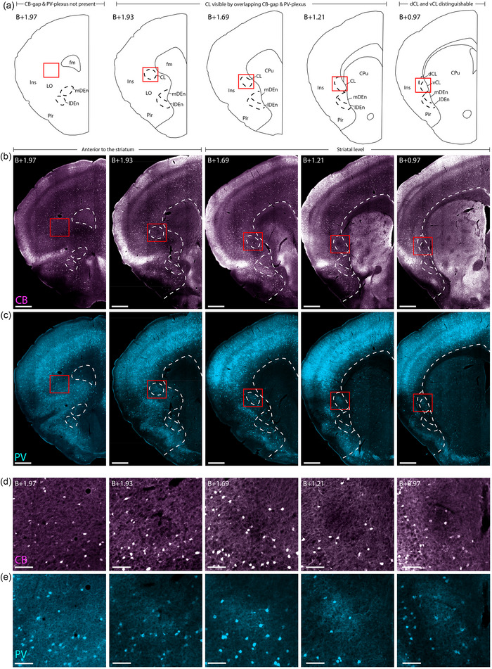
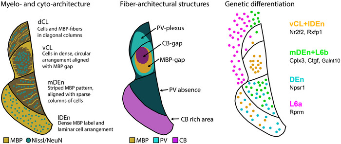
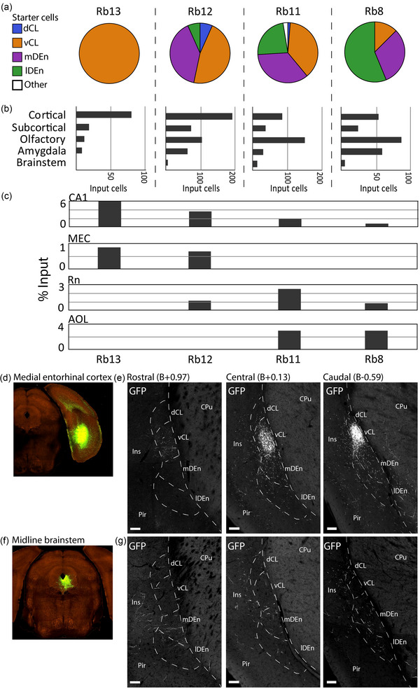
Accurate anatomical characterizations are necessary to investigate neural circuitry on a fine scale, but for the rodent claustrum complex (CLCX), this has yet to be fully accomplished. The CLCX is generally considered to comprise two major subdivisions, the claustrum (CL) and the dorsal endopiriform nucleus (DEn), but regional boundaries to these areas are debated. To address this, we conducted a multifaceted analysis of fiber- and cytoarchitecture, genetic marker expression, and connectivity using mice of both sexes, to create a comprehensive guide for identifying and delineating borders to CLCX, including an online reference atlas. Our data indicated four distinct subregions within CLCX, subdividing both CL and DEn into two. Additionally, we conducted brain-wide tracing of inputs to CLCX using a transgenic mouse line. Immunohistochemical staining against myelin basic protein (MBP), parvalbumin (PV), and calbindin (CB) revealed intricate fiber-architectural patterns enabling precise delineations of CLCX and its subregions. Myelinated fibers were abundant dorsally in CL but absent ventrally, whereas PV expressing fibers occupied the entire CL. CB staining revealed a central gap within CL, also visible anterior to the striatum. The Nr2f2, Npsr1, and Cplx3 genes expressed specifically within different subregions of the CLCX, and Rprm helped delineate the CL-insular border. Furthermore, cells in CL projecting to the retrosplenial cortex were located within the myelin sparse area. By combining own experimental data with digitally available datasets of gene expression and input connectivity, we could demonstrate that the proposed delineation scheme allows anchoring of datasets from different origins to a common reference framework.
精确的解剖学特征描述对于在精细尺度上研究神经回路是必要的,但对于啮齿动物的屏状核复合体(CLCX)而言,这一点尚未完全实现。CLCX通常被认为由两个主要亚区组成,即屏状核(CL)和背侧内梨状核(DEn),但这些区域的边界存在争议。为了解决这个问题,我们使用雌雄小鼠对纤维结构、细胞结构、遗传标记表达和连接性进行了多方面分析,以创建一份全面的指南,用于识别和描绘CLCX的边界,包括一个在线参考图谱。我们的数据表明CLCX内有四个不同的亚区,将CL和DEn都细分为两个部分。此外,我们使用转基因小鼠品系对输入到CLCX的脑区进行了全脑追踪。针对髓鞘碱性蛋白(MBP)、小白蛋白(PV)和钙结合蛋白(CB)的免疫组织化学染色揭示了复杂的纤维结构模式,能够精确描绘CLCX及其亚区。CL的背侧有丰富的有髓纤维,但腹侧没有,而表达PV的纤维占据了整个CL。CB染色显示CL内有一个中央间隙,在纹状体前方也可见。Nr2f2、Npsr1和Cplx3基因在CLCX的不同亚区内特异性表达,Rprm有助于描绘CL与岛叶的边界。此外,投射到 retrosplenial 皮质的CL中的细胞位于髓鞘稀疏区域内。通过将我们自己的实验数据与数字可得的基因表达和输入连接性数据集相结合,我们可以证明所提出的描绘方案能够将来自不同来源的数据集锚定到一个共同的参考框架。