Meikle P J, Perry M B, Cherwonogrodzky J W, Bundle D R
Division of Biological Sciences, National Research Council of Canada, Ottawa, Ontario.
Infect Immun. 1989 Sep;57(9):2820-8. doi: 10.1128/iai.57.9.2820-2828.1989.
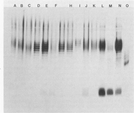
Brucella A and M epitopes were found on single O-polysaccharide chains of all biotype strains of this species. Lipopolysaccharides from the type and reference strains of five of the six Brucella species, B. abortus, B. melitensis, B. suis, B. canis, and B. neotomae, were extracted and purified. Analysis by sodium dodecyl sulfate-polyacrylamide gel electrophoresis, in conjunction with silver staining and immunoblotting developed by monoclonal antibodies, showed bands characteristic of A, M, or mixed A and M antigens. The A antigen previously described as an exclusively alpha 1,2-linked homopolymer of 4,6-dideoxy-4-formamido-D-mannopyranose was shown by 1H and 13C nuclear magnetic resonance spectroscopy to possess a fine structure consistent with the low-frequency occurrence of alpha 1, 3-linked 4,6-dideoxy-4-formamido-D-mannopyranose residues. This feature was previously attributed only to the M antigen, which is also a homopolymer of the same sugar. B. melitensis biotype 3 and B. suis biotype 4 lipopolysaccharides showed characteristics of mixed A and M antigens. Immunoabsorption of these O polysaccharides on a column of immobilized A-antigen-specific monoclonal antibody enriched polymer chains with A-antigen characteristics but did not eliminate M epitopes. Composite A- and M-antigen characteristics resulted from O polysaccharides in which the frequency of alpha 1,3 linkages, and hence, M-antigen characteristics, varied. All biotypes assigned as A+ M- expressed one or two alpha 1,3-linked residues per polysaccharide O chain. M antigens (M+ A-) also possessed a unique M epitope as well as a tetrasaccharide determinant common to A-antigen structures. B. canis and B. abortus 45/20, both rough strains, expressed low-molecular-weight A antigen.
在该物种所有生物型菌株的单个O-多糖链上发现了布鲁氏菌A和M表位。提取并纯化了六种布鲁氏菌中的五种——流产布鲁氏菌、羊布鲁氏菌、猪布鲁氏菌、犬布鲁氏菌和新墨西哥布鲁氏菌的模式菌株和参考菌株的脂多糖。通过十二烷基硫酸钠-聚丙烯酰胺凝胶电泳分析,结合银染和单克隆抗体免疫印迹法,显示出A、M或A和M混合抗原的特征条带。先前被描述为4,6-二脱氧-4-甲酰胺基-D-甘露吡喃糖的仅α1,2连接的均聚物的A抗原,通过1H和13C核磁共振光谱显示其精细结构与α1,3连接的4,6-二脱氧-4-甲酰胺基-D-甘露吡喃糖残基的低频出现一致。该特征先前仅归因于M抗原,M抗原也是相同糖的均聚物。羊布鲁氏菌生物型3和猪布鲁氏菌生物型4的脂多糖表现出A和M混合抗原的特征。这些O多糖在固定化A抗原特异性单克隆抗体柱上的免疫吸附富集了具有A抗原特征的聚合物链,但未消除M表位。复合A和M抗原特征是由α1,3连接频率以及因此M抗原特征不同的O多糖产生的。所有被指定为A + M-的生物型每条多糖O链表达一个或两个α1,3连接的残基。M抗原(M + A-)还具有独特M表位以及A抗原结构共有的四糖决定簇。犬布鲁氏菌和流产布鲁氏菌45/20这两种粗糙菌株均表达低分子量A抗原。