Kawasaki Takayasu, Fujioka Jun, Imai Takayuki, Torigoe Kanjiro, Tsukiyama Koichi
IR Free Electron Laser Research Center, Research Institute for Science and Technology (RIST), Tokyo University of Science, 2641, Yamazaki, Noda, Chiba, 278-8510, Japan,
Lasers Med Sci. 2014 Sep;29(5):1701-7. doi: 10.1007/s10103-014-1577-5. Epub 2014 Apr 24.
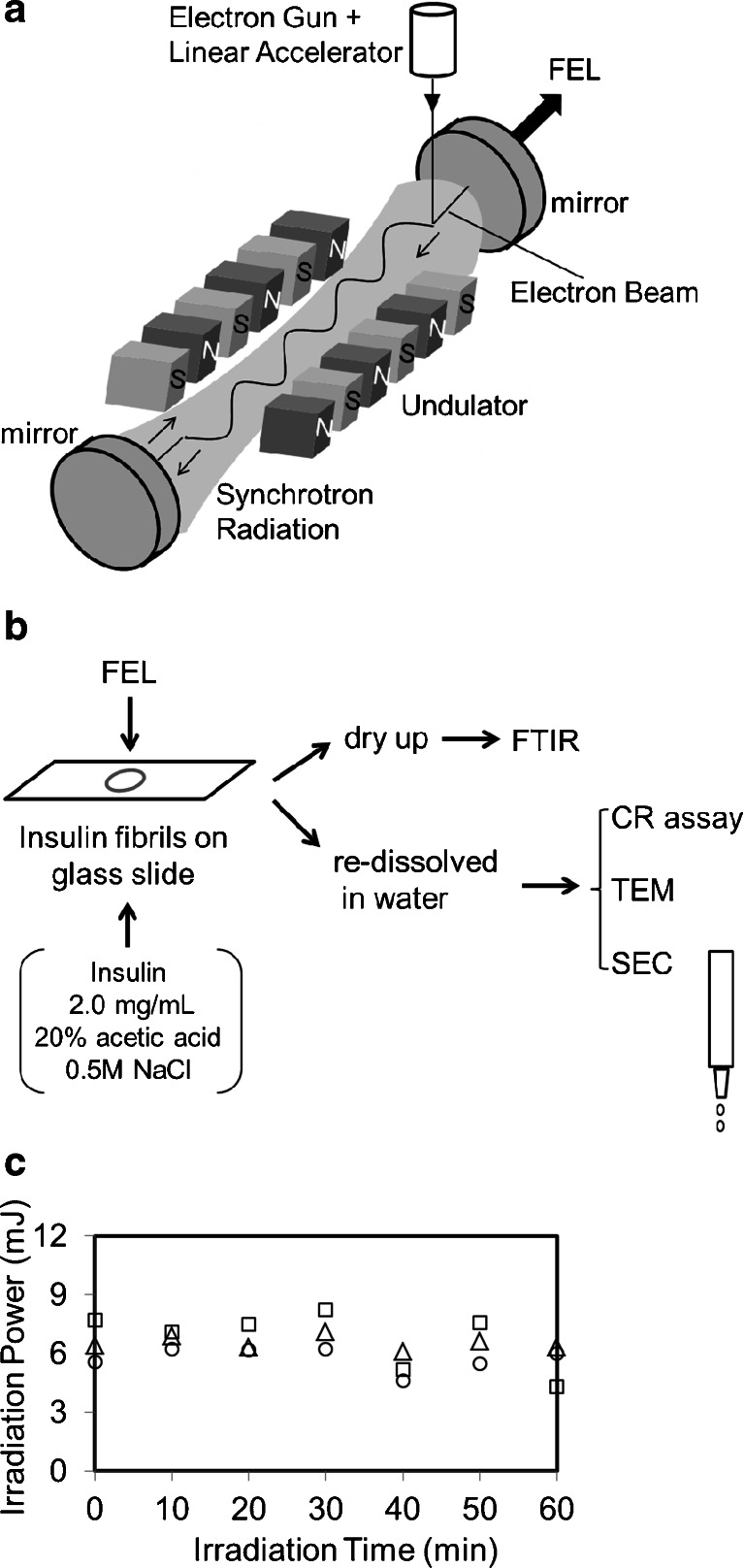
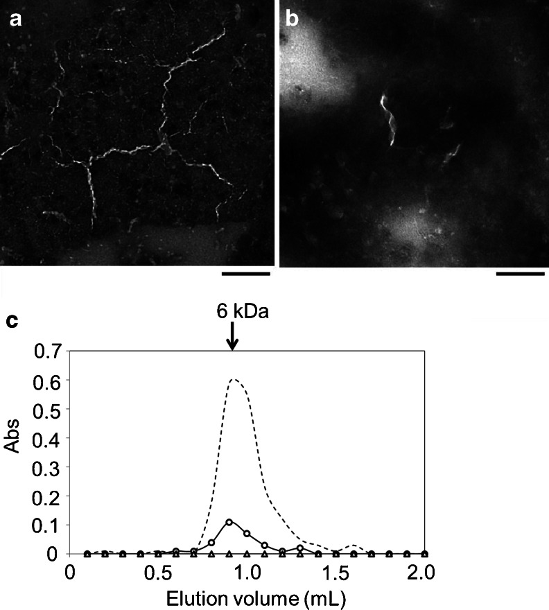
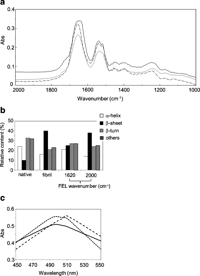
A mid-infrared free-electron laser (FEL) is operated as a pulsed and linearly polarized laser with tunable wavelengths within infrared region. Although the FEL can ablate soft tissues with minimum collateral damage in surgery, the potential of FEL for dissecting protein aggregates is not fully understood. Protein aggregates such as amyloid fibrils are in some cases involved in serious diseases. In our previous study, we showed that amyloid-like lysozyme fibrils could be disaggregated into the native form with FEL irradiation specifically tuned to the amide I band (1,620 cm(-1)). Here, we show further evidence for the FEL-mediated disaggregation of amyloid-like fibrils using insulin fibrils. Insulin fibrils were prepared in acidic solution and irradiated by the FEL, which was tuned to either 1,620 or 2,000 cm(-1) prior to the experiment. The Fourier transform infrared spectroscopy (FT-IR) spectrum after irradiation with the FEL at 1,620 cm(-1) indicated that the broad peak (1,630-1,660 cm(-1)) became almost a single peak (1,652 cm(-1)), and the β-sheet content was reduced to 25 from 40% in the fibrils, while that following the irradiation at 2,000 cm(-1) remained at 38%. The Congo Red assay as well as transmission electron microscopy observation confirmed that the number of fibrils was reduced by FEL irradiation at the amide I band. Size-exclusion chromatography analysis indicated that the disaggregated form of fibrils was the monomeric form. These results confirm that FEL irradiation at the amide I band can dissect amyloid-like protein fibrils into the monomeric form in vitro.
中红外自由电子激光(FEL)作为一种脉冲且线偏振的激光运行,其波长在红外区域内可调。尽管FEL在手术中能够以最小的附带损伤消融软组织,但其用于分解蛋白质聚集体的潜力尚未得到充分了解。诸如淀粉样原纤维等蛋白质聚集体在某些情况下与严重疾病有关。在我们之前的研究中,我们表明,通过将FEL辐射专门调谐至酰胺I带(1620 cm⁻¹),淀粉样溶菌酶原纤维可以解聚为天然形式。在这里,我们使用胰岛素原纤维展示了FEL介导的淀粉样原纤维解聚的进一步证据。胰岛素原纤维在酸性溶液中制备,并在实验前用调谐至1620或2000 cm⁻¹的FEL进行照射。在1620 cm⁻¹处用FEL照射后的傅里叶变换红外光谱(FT-IR)表明,宽峰(1630 - 1660 cm⁻¹)几乎变成了单峰(1652 cm⁻¹),并且原纤维中的β-折叠含量从40%降至25%,而在2000 cm⁻¹处照射后的β-折叠含量保持在38%。刚果红测定以及透射电子显微镜观察证实,在酰胺I带处用FEL照射会减少原纤维的数量。尺寸排阻色谱分析表明,解聚后的原纤维形式为单体形式。这些结果证实,在酰胺I带处用FEL照射可以在体外将淀粉样蛋白原纤维分解为单体形式。