Kumral Tolgar Lütfi, Yildirim Güven, Uyar Yavuz
2 Department of Otolaryngology, Okmeydani Training and Research Hospital, Istanbul, Turkey.
Clin Pract. 2012 Jan 9;2(1):e10. doi: 10.4081/cp.2012.e10. eCollection 2012 Jan 1.
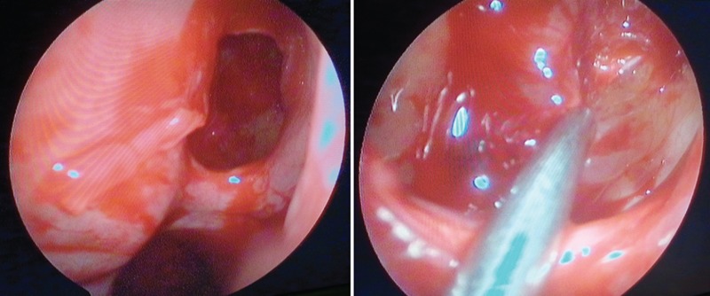
Isolated sphenoid pathology is uncommon. Nasal polyps that originate from the anterior wall of the sphenoid sinus and reach the nasopharynx are called sphenochoanal polyps. The atypical location of sphenochoanal polyps leads to misdiagnosis, and surgery risks injuring the surrounding structures, such as the optic nerve, carotid artery, and brain. For the differential diagnosis of sphenochoanal polyps, nasal endoscopy and computed tomography are very important. We present the clinical and radiological features of a sphenochoanal polyp and review the status of the optic nerve during endoscopic surgery for a sphenochoanal polyp.
孤立性蝶窦病变并不常见。起源于蝶窦前壁并延伸至鼻咽部的鼻息肉称为蝶筛隐窝息肉。蝶筛隐窝息肉的非典型位置会导致误诊,手术有损伤周围结构的风险,如视神经、颈动脉和脑。对于蝶筛隐窝息肉的鉴别诊断,鼻内镜检查和计算机断层扫描非常重要。我们介绍了一例蝶筛隐窝息肉的临床和影像学特征,并回顾了蝶筛隐窝息肉内镜手术中视神经的情况。