Department of Breast Oncology, International Medical Center, Saitama Medical University, Hidaka, Saitama.
Cancer Sci. 2014 Jul;105(7):833-9. doi: 10.1111/cas.12432. Epub 2014 Jun 27.
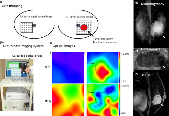
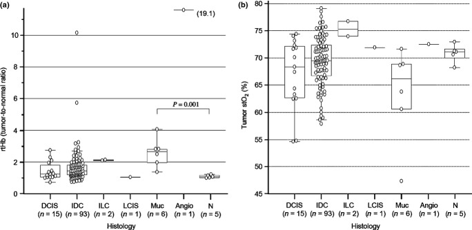
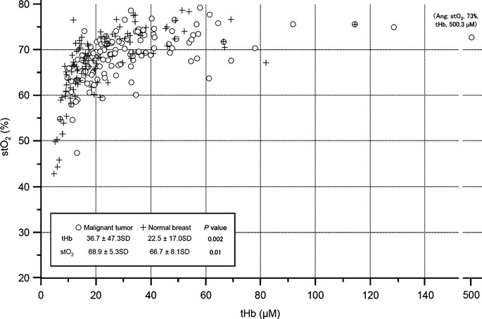
Near-infrared diffuse optical spectroscopy (DOS) imaging can non-invasively measure tumor hemoglobin concentration using high contrast to normal tissue, thus providing vascularity and oxygenation status. We assessed the clinical usefulness of DOS imaging in primary breast cancer. In all, 118 women with a histologically confirmed diagnosis of primary malignant tumor were enrolled. All participants underwent testing using time-resolved DOS before treatment initiation. Visual assessment of DOS imaging for detecting tumors was carried out by two readers blinded to the clinical data. Relative total hemoglobin (rtHb) and oxygen saturation (stO2 ) of the tumors was compared with clinicopathological variables and 10-year prognosis was calculated. Sensitivity for detecting a tumor based on the rtHb breast map was 62.7% (74/118). The sensitivity depended on T stage: 100% (7/7) for T3, 78.9% (45/57) for T2, 44.7% (17/38) for T1, and 31.3% (5/16) for Tis . Tumors showed unique features of higher rtHb with a wider range of stO2 than normal breast tissue, depending on histological type. There was a significant correlation of rtHb with tumor size, lymphatic vascular invasion, and histological grade, and of stO2 with age and tumor size. Neither rtHb nor stO2 correlated with intrinsic biomarkers such as estrogen receptor, progesterone receptor, or human epidermal growth factor receptor 2; rtHb inversely correlated with 10-year relapse-free survival and overall survival, with statistical significance. Diffuse optical spectroscopy imaging has limited utility for the early detection of breast cancer; nonetheless, the findings suggest that the degree of tumor angiogenesis and hypoxia may be associated with tumor aggressiveness and poor prognosis.
近红外漫射光学光谱(DOS)成像是一种非侵入性的方法,可以通过高对比度来测量肿瘤的血红蛋白浓度,从而提供血管生成和氧合状态。我们评估了 DOS 成像在原发性乳腺癌中的临床应用价值。共纳入 118 例经组织学证实为原发性恶性肿瘤的女性患者。所有患者在治疗前均进行了时间分辨 DOS 检测。两名对临床数据不知情的读者对 DOS 成像检测肿瘤的能力进行了视觉评估。比较了肿瘤的相对总血红蛋白(rtHb)和氧饱和度(stO2)与临床病理变量的关系,并计算了 10 年的预后。基于 rtHb 乳腺图检测肿瘤的敏感性为 62.7%(74/118)。敏感性取决于 T 分期:T3 期为 100%(7/7),T2 期为 78.9%(45/57),T1 期为 44.7%(17/38),Tis 期为 31.3%(5/16)。根据组织学类型,肿瘤表现出独特的特征,即 rtHb 较高,stO2 范围较宽。rtHb 与肿瘤大小、淋巴管血管侵犯和组织学分级显著相关,stO2 与年龄和肿瘤大小显著相关。rtHb 与雌激素受体、孕激素受体或人表皮生长因子受体 2 等内在生物标志物均无相关性,stO2 与年龄和肿瘤大小显著相关。rtHb 与 10 年无复发生存率和总生存率呈负相关,具有统计学意义。漫射光学光谱成像在早期检测乳腺癌方面的应用价值有限;然而,研究结果表明,肿瘤血管生成和缺氧程度可能与肿瘤侵袭性和不良预后相关。