Hosen Mohammad J, Coucke Paul J, Le Saux Olivier, De Paepe Anne, Vanakker Olivier M
Center for Medical Genetics, Ghent University Hospital, Ghent, Belgium.
Orphanet J Rare Dis. 2014 Apr 29;9:66. doi: 10.1186/1750-1172-9-66.
Pseudoxanthoma elasticum (PXE) is characterized by skin (papular lesions), ocular (subretinal neovascularisation) and cardiovascular manifestations (peripheral artery disease), due to mineralization and fragmentation of elastic fibres in the extracellular matrix (ECM). Caused by mutations in the ABCC6 gene, the mechanisms underlying this disease remain unknown. The knowledge on the molecular background of soft tissue mineralization largely comes from insights in vascular calcification, with involvement of the osteoinductive Transforming Growth Factor beta (TGFβ) family (TGFβ1-3 and Bone Morphogenetic Proteins [BMP]), together with ectonucleotides (ENPP1), Wnt signalling and a variety of local and systemic calcification inhibitors. In this study, we have investigated the relevance of the signalling pathways described in vascular soft tissue mineralization in the PXE knock-out mouse model and in PXE patients.
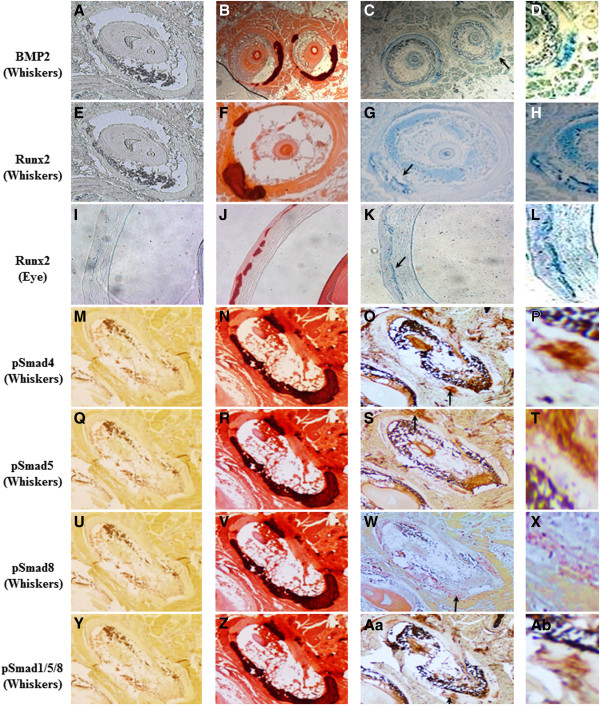
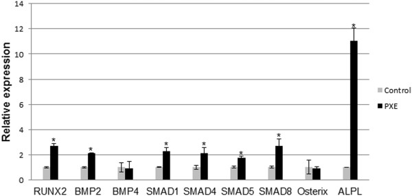
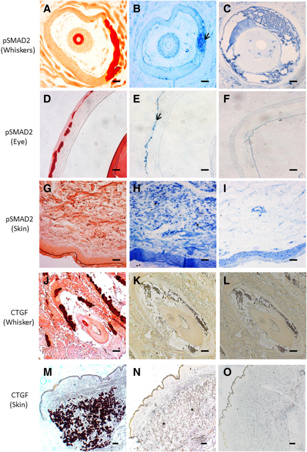
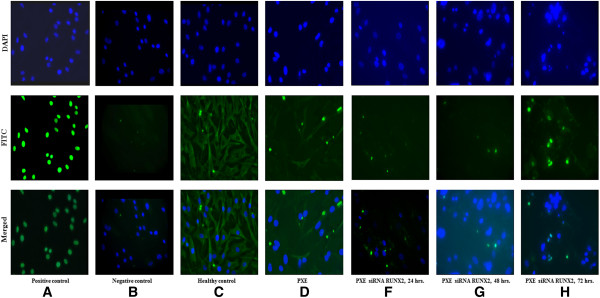
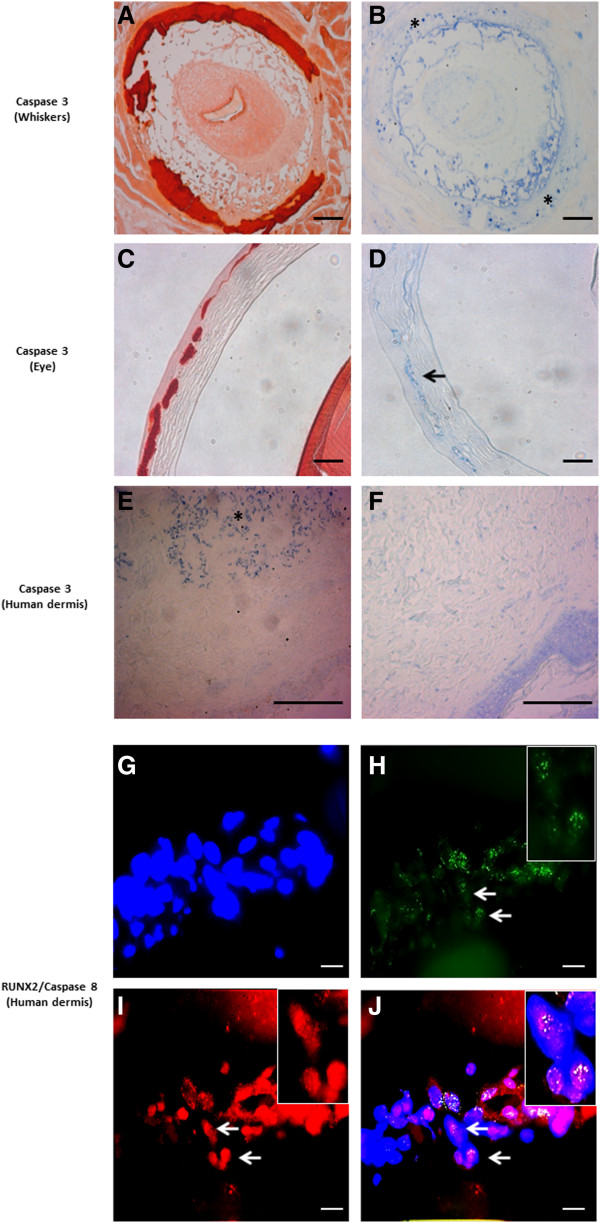
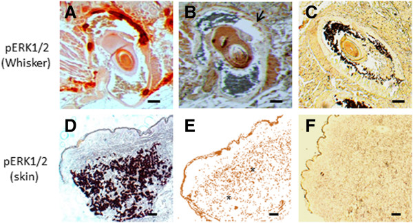
The role of the pro-osteogenic pathways BMP2-SMADs-RUNX2, TGFβ-SMAD2/3 and Wnt-MSX2, apoptosis and ER stress was evaluated using immunohistochemistry, mRNA expression profiling and immune-co-staining in dermal tissues and fibroblast cultures of PXE patients and the eyes and whiskers of the PXE knock-out mouse. Apoptosis was further evaluated by TUNEL staining and siRNA mediated gene knockdown. ALPL activity in PXE fibroblasts was studied using ALPL stains.
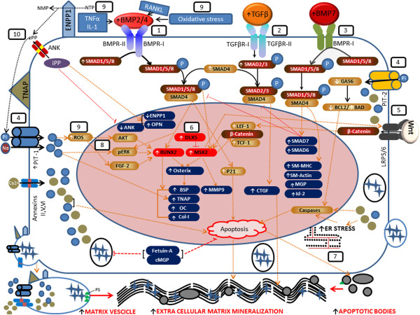
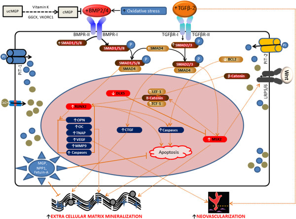
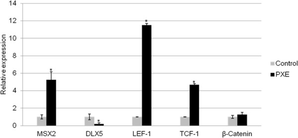
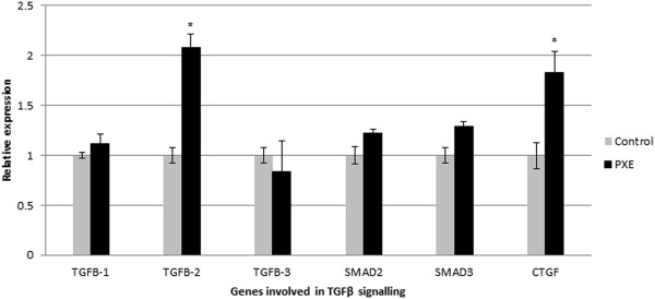
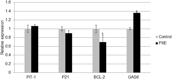
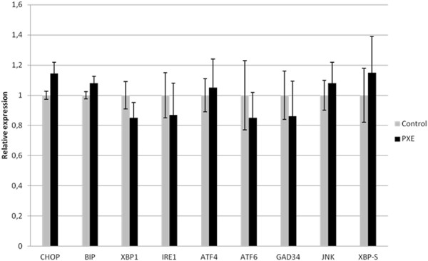
We demonstrate the upregulation of the BMP2-SMADs-RUNX2 and TGFβ-2-SMAD2/3 pathway, co-localizing with the mineralization sites, and the involvement of MSX2-canonical Wnt signalling. Further, we show that apoptosis is also involved in PXE with activation of Caspases and BCL-2. In contrast to vascular calcification, neither the other BMPs and TGFβs nor endoplasmic reticulum stress pathways seem to be perturbed in PXE.
Our study shows that we cannot simply extrapolate knowledge on cell signalling in vascular soft tissue calcification to a multisystem ectopic mineralisation disease as PXE. Contrary, we demonstrate a specific set of perturbed signalling pathways in PXE patients and the knock-out mouse model. Based on our findings and previously reported data, we propose a preliminary cell model of ECM calcification in PXE.
弹性假黄瘤(PXE)的特征在于皮肤(丘疹性病变)、眼部(视网膜下新生血管形成)和心血管表现(外周动脉疾病),这是由于细胞外基质(ECM)中弹性纤维的矿化和断裂所致。该疾病由ABCC6基因突变引起,但其潜在机制仍不清楚。关于软组织矿化分子背景的知识主要来自对血管钙化的研究,涉及骨诱导性转化生长因子β(TGFβ)家族(TGFβ1 - 3和骨形态发生蛋白[BMP]),以及外核苷酸(ENPP1)、Wnt信号通路和多种局部及全身钙化抑制剂。在本研究中,我们调查了在PXE基因敲除小鼠模型和PXE患者中,血管软组织矿化中描述的信号通路的相关性。
使用免疫组织化学、mRNA表达谱分析和免疫共染色,在PXE患者的皮肤组织和成纤维细胞培养物以及PXE基因敲除小鼠的眼睛和触须中,评估促骨生成通路BMP2 - SMADs - RUNX2、TGFβ - SMAD2/3和Wnt - MSX2、细胞凋亡和内质网应激的作用。通过TUNEL染色和siRNA介导的基因敲低进一步评估细胞凋亡。使用ALPL染色研究PXE成纤维细胞中的ALPL活性。
我们证明了BMP2 - SMADs - RUNX2和TGFβ - 2 - SMAD2/3通路的上调,它们与矿化位点共定位,以及MSX2 - 经典Wnt信号通路的参与。此外,我们表明细胞凋亡也参与了PXE,伴有半胱天冬酶和BCL - 2的激活。与血管钙化不同,其他BMP和TGFβ以及内质网应激通路在PXE中似乎未受干扰。
我们的研究表明,我们不能简单地将血管软组织钙化中的细胞信号知识外推到像PXE这样的多系统异位矿化疾病中。相反,我们在PXE患者和基因敲除小鼠模型中证明了一组特定的受干扰信号通路。基于我们的发现和先前报道的数据,我们提出了PXE中ECM钙化的初步细胞模型。