Crapanzano John P, Heymann Jonas J, Monaco Sara, Nassar Aziza, Saqi Anjali
Address: Department of Pathology and Cell Biology, Columbia University Medical Center, New York-Presbyterian Hospital, New York, NY, USA.
University of Pittsburgh Medical Center, Pittsburgh, PA, USA.
Cytojournal. 2014 Mar 20;11:7. doi: 10.4103/1742-6413.129187. eCollection 2014.
In the recent past, algorithms and recommendations to standardize the morphological, immunohistochemical and molecular classification of lung cancers on cytology specimens have been proposed, and several organizations have recommended cell blocks (CBs) as the preferred modality for molecular testing. Based on the literature, there are several different techniques available for CB preparation-suggesting that there is no standard. The aim of this study was to conduct a survey of CB preparation techniques utilized in various practice settings and analyze current issues, if any.
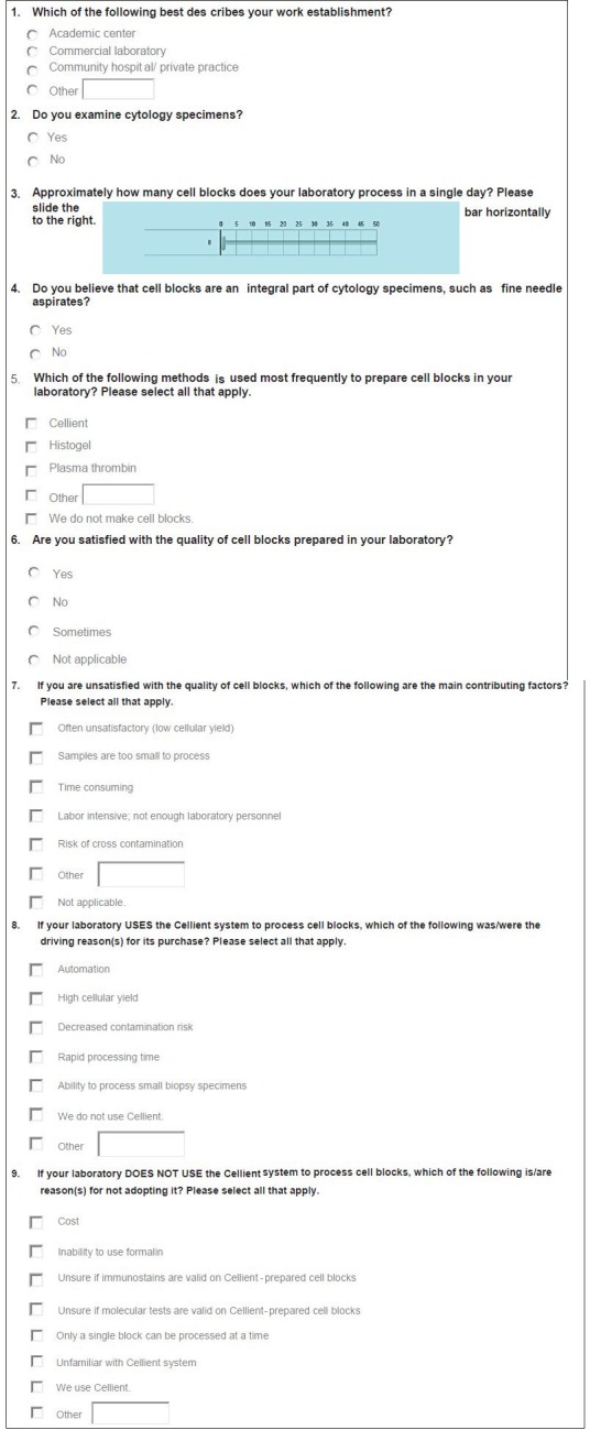
A single E-mail with a link to an electronic survey was distributed to members of the American Society of Cytopathology and other pathologists. Questions pertaining to the participants' practice setting and CBs-volume, method, quality and satisfaction-were included.
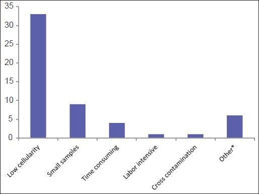
Of 95 respondents, 90/95 (94%) completed the survey and comprise the study group. Most participants practice in a community hospital/private practice (44%) or academic center (41%). On average, 14 CBs (range 0-50; median 10) are prepared by a laboratory daily. Over 10 methods are utilized: Plasma thrombin (33%), HistoGel (27%), Cellient automated cell block system (8%) and others (31%) respectively. Forty of 90 (44%) respondents are either unsatisfied or sometimes satisfied with their CB quality, with low-cellular yield being the leading cause of dissatisfaction. There was no statistical significance between the three most common CB preparation methods and satisfaction with quality.
Many are dissatisfied with their current method of CB preparation, and there is no consistent method to prepare CBs. In today's era of personalized medicine with an increasing array of molecular tests being applied to cytological specimens, there is a need for a standardized protocol for CB optimization to enhance cellularity.
最近,有人提出了使肺癌细胞学标本的形态学、免疫组织化学及分子分类标准化的算法和建议,一些组织推荐使用细胞块(CBs)作为分子检测的首选方式。根据文献,有几种不同的细胞块制备技术,这表明尚无标准方法。本研究的目的是对各种实际应用场景中使用的细胞块制备技术进行调查,并分析当前存在的问题(如有)。
向美国细胞病理学会成员及其他病理学家发送了一封包含电子调查问卷链接的电子邮件。问卷内容包括与参与者的实际应用场景以及细胞块的数量、制备方法、质量和满意度相关的问题。
95名受访者中,90/95(94%)完成了调查,构成研究组。大多数参与者就职于社区医院/私人诊所(44%)或学术中心(41%)。实验室每天平均制备14个细胞块(范围0 - 50;中位数10)。使用的方法超过10种:分别为血浆凝血酶(33%)、组织胶(27%)、Cellient自动细胞块系统(8%)和其他方法(31%)。90名受访者中有40名(44%)对其细胞块质量不满意或有时满意,细胞产量低是导致不满的主要原因。三种最常用的细胞块制备方法与质量满意度之间无统计学差异。
许多人对他们目前的细胞块制备方法不满意,且没有一致的细胞块制备方法。在当今个性化医疗时代,越来越多的分子检测应用于细胞学标本,需要一种标准化方案来优化细胞块以提高细胞含量。