Zou Yimin, Li Shaoxing, Zou Weifeng, Hu Guoping, Zhou Yumin, Peng Gongyong, He Fang, Li Bing, Ran Pixin
The State Key Laboratory of Respiratory Disease, Guangzhou Institute of Respiratory Diseases, the First Affiliated Hospital, Guangzhou Medical University, Guangzhou, Guangdong, China.
Guangzhou Chest Hospital, Guangzhou Medical University, Guangzhou, Guangdong, China.
PLoS One. 2014 May 6;9(5):e96708. doi: 10.1371/journal.pone.0096708. eCollection 2014.
Peribronchiolar fibrosis is an important feature of small airway remodeling (SAR) in cigarette smoke-induced COPD. The aim of this study was to investigate the role of gelatinases (MMP9, MMP2) and epithelial-mesenchymal transition (EMT) in SAR related to wood smoke (WS) exposure in a rat model.
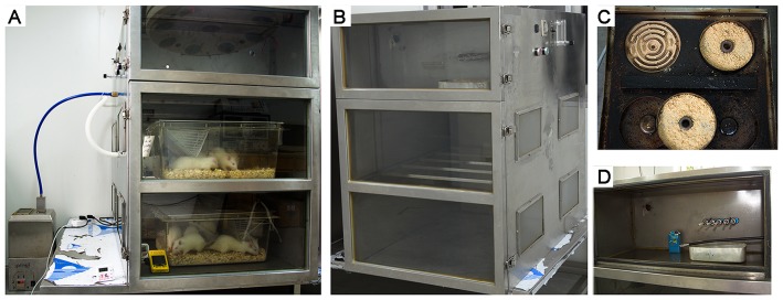
Forty-eight female Sprague-Dawley rats were randomly divided into the WS group, the cigarette smoke (CS) group and the clean air control group. After 4 to 7 months of smoke exposure, lung tissues were examined with morphometric measurements, immunohistochemistry and Western blotting. Serum MMP9 and TIMP1 concentrations were detected by ELISA. In vitro, primary rat tracheal epithelial cells were stimulated with wood smoke condensate for 7 days.
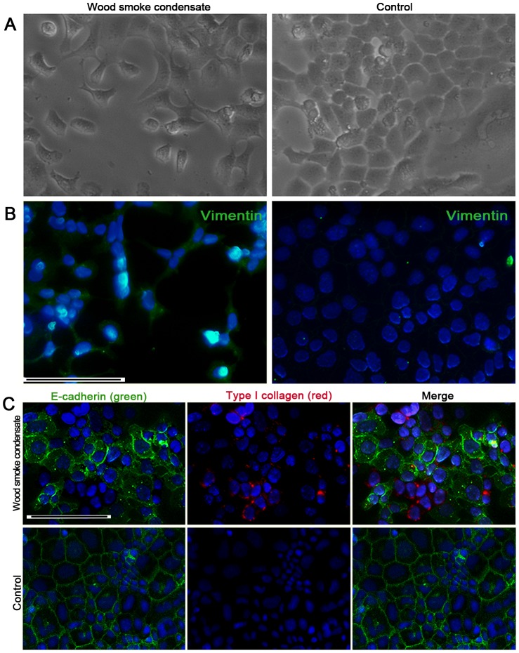
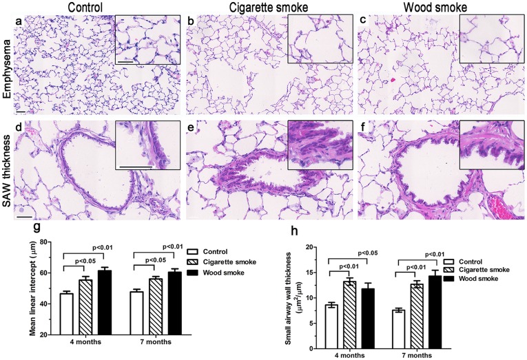
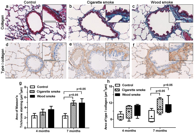
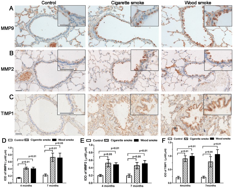
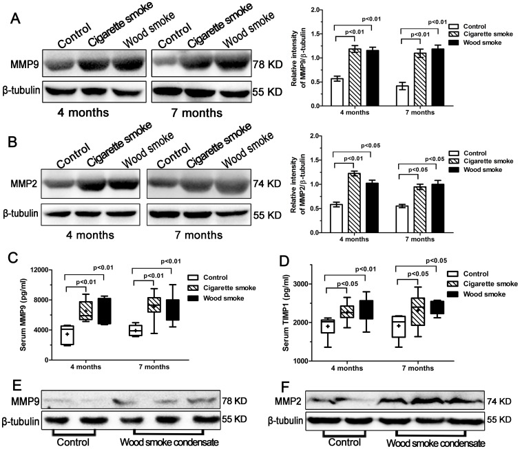
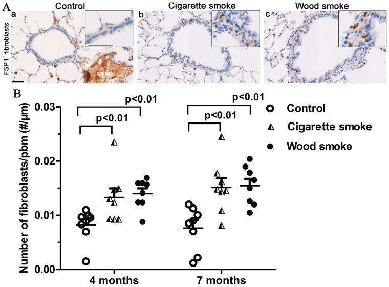
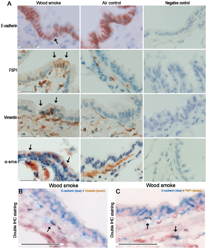
The COPD-like pathological alterations in rats exposed chronically to WS were similar to those exposed to CS; the area of collagen deposition was significantly increased in the small airway walls of those exposed to WS or CS for 7 months. The expression of gelatinases in rats induced by WS or CS exposure was markedly increased in whole lung tissue, and immunohistochemistry showed that MMP9, MMP2 and TIMP1 were primarily expressed in the airway epithelium. The serum levels of MMP9 and TIMP1 were significantly higher in rats secondary to WS or CS exposure. Few cells that double immunostained for E-cadherin and vimentin were observed in the airway subepithelium of rats exposed to WS for 7 months (only 3 of these 8 rats). In vitro, the expression of MMP9 and MMP2 proteins was upregulated in primary rat tracheal epithelial cells following exposure to wood smoke condensate for 7 days by Western blotting; positive immunofluorescent staining for vimentin and type I collagen was also observed.
These findings suggest that the upregulation of gelatinases and EMT might play a role in SAR in COPD associated with chronic exposure to wood smoke.
细支气管周围纤维化是香烟烟雾诱导的慢性阻塞性肺疾病(COPD)中小气道重塑(SAR)的一个重要特征。本研究旨在探讨明胶酶(基质金属蛋白酶9、基质金属蛋白酶2)和上皮-间质转化(EMT)在与木烟(WS)暴露相关的大鼠模型SAR中的作用。
48只雌性Sprague-Dawley大鼠随机分为WS组、香烟烟雾(CS)组和清洁空气对照组。经过4至7个月的烟雾暴露后,通过形态计量学测量、免疫组织化学和蛋白质印迹法对肺组织进行检查。采用酶联免疫吸附测定法检测血清基质金属蛋白酶9和金属蛋白酶组织抑制因子1的浓度。在体外,用木烟冷凝物刺激原代大鼠气管上皮细胞7天。
长期暴露于WS的大鼠出现的类似COPD的病理改变与暴露于CS的大鼠相似;暴露于WS或CS 7个月的大鼠小气道壁中胶原沉积面积显著增加。WS或CS暴露诱导的大鼠全肺组织中明胶酶的表达明显增加,免疫组织化学显示基质金属蛋白酶9、基质金属蛋白酶2和金属蛋白酶组织抑制因子1主要在气道上皮中表达。暴露于WS或CS的大鼠血清基质金属蛋白酶9和金属蛋白酶组织抑制因子1水平显著升高。暴露于WS 7个月的大鼠气道上皮下很少观察到E-钙黏蛋白和波形蛋白双重免疫染色的细胞(这8只大鼠中只有3只)。在体外,通过蛋白质印迹法检测发现,原代大鼠气管上皮细胞暴露于木烟冷凝物7天后,基质金属蛋白酶9和基质金属蛋白酶2蛋白的表达上调;同时也观察到波形蛋白和I型胶原的阳性免疫荧光染色。
这些发现表明,明胶酶的上调和EMT可能在与长期暴露于木烟相关的COPD的SAR中起作用。