• 文献检索
  • 文档翻译
  • 深度研究
  • 学术资讯
  • Suppr Zotero 插件Zotero 插件
  • 邀请有礼
  • 套餐&价格
  • 历史记录
应用&插件
Suppr Zotero 插件Zotero 插件浏览器插件Mac 客户端Windows 客户端微信小程序
定价
高级版会员购买积分包购买API积分包
服务
文献检索文档翻译深度研究API 文档MCP 服务
关于我们
关于 Suppr公司介绍联系我们用户协议隐私条款
关注我们

Suppr 超能文献

核心技术专利:CN118964589B侵权必究
粤ICP备2023148730 号-1Suppr @ 2026

文献检索

告别复杂PubMed语法,用中文像聊天一样搜索,搜遍4000万医学文献。AI智能推荐,让科研检索更轻松。

立即免费搜索

文件翻译

保留排版,准确专业,支持PDF/Word/PPT等文件格式,支持 12+语言互译。

免费翻译文档

深度研究

AI帮你快速写综述,25分钟生成高质量综述,智能提取关键信息,辅助科研写作。

立即免费体验

胎儿期人类小脑叶外表面的发育

Development of external surfaces of human cerebellar lobes in the fetal period.

作者信息

Nowakowska-Kotas Marta, Kędzia Alicja, Dudek Krzysztof

机构信息

Department of Neurology, Wrocław Medical University, ul. Borowska 213, 50-556, Wrocław, Poland,

出版信息

Cerebellum. 2014 Oct;13(5):541-8. doi: 10.1007/s12311-014-0566-3.

DOI:10.1007/s12311-014-0566-3
PMID:24831768
原文链接:https://pmc.ncbi.nlm.nih.gov/articles/PMC4155164/
Abstract

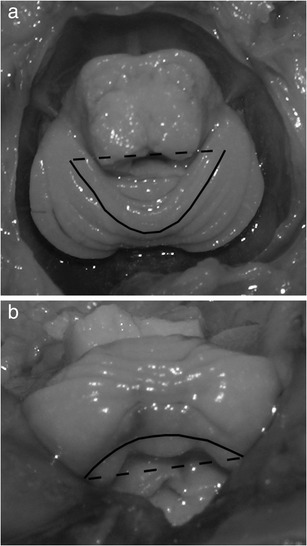
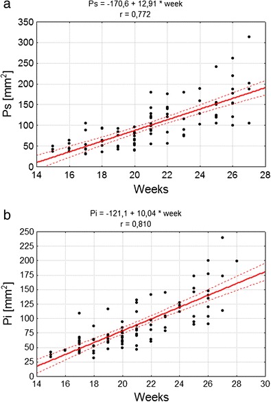
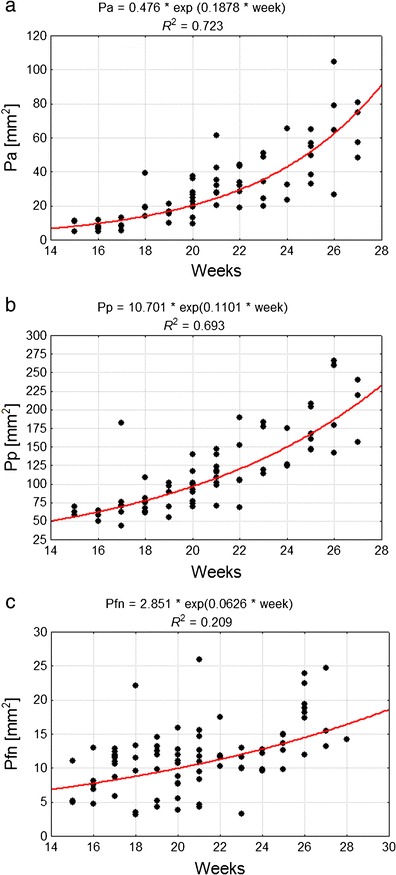
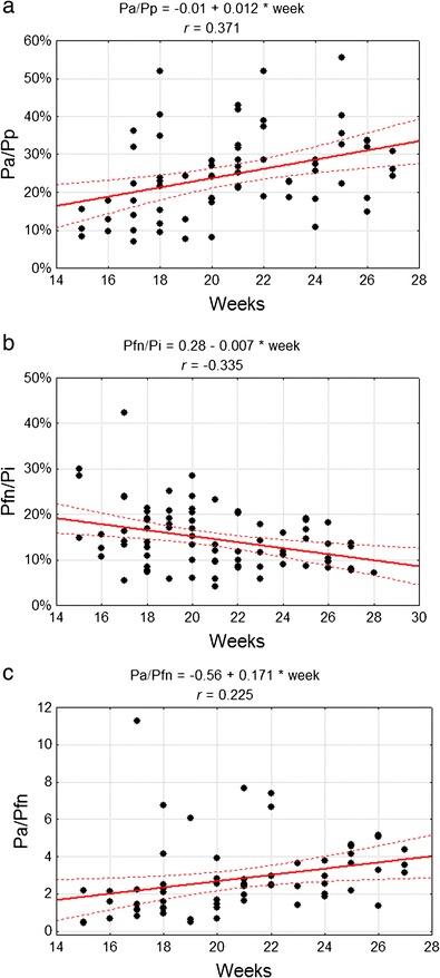
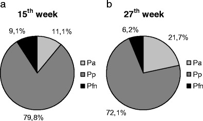
In the fetal period, development of cerebellar lobes may proceed dissimilarly due to possible differentiated origins of the cells and diversified times of their migration to certain cerebellum regions. This can cause various growth trajectories for the external surfaces of cerebellar lobes. The goal of the study was to describe the development of the external surface of cerebellum lobes and fissures delineating them in the fetal period. The material consisted of 101 fetuses (48 males and 53 females)-crown rump length 89-229 mm corresponding to 15-28 weeks of fetal life. The methods were based on anthropometric measurements and preparation techniques combined with elicited image computer analysis. At the largest values of the cerebellum posterior lobe surface, the most dynamic growth rate was observed in the case of the anterior lobe. Among the cerebellar lobes, proportional change was observed as well as a gradual increase in anterior lobe surface area and a simultaneous decrease in the surface area of the flocculonodular lobe part of the cerebellum total external surface. This paper presents the different growth trajectories of cerebellar lobes and demonstrates the importance of the primary fissure as a delineating mark for two regions with different dynamics of development.

摘要

在胎儿期,小脑叶的发育可能会因细胞起源的差异以及它们迁移到特定小脑区域的时间不同而有所不同。这可能导致小脑叶外表面出现各种生长轨迹。本研究的目的是描述胎儿期小脑叶外表面及其裂隙的发育情况。研究材料包括101例胎儿(48例男性和53例女性),顶臀长89 - 229毫米,相当于胎儿15 - 28周的孕期。研究方法基于人体测量学测量和制备技术,并结合图像计算机分析。在小脑后叶表面面积最大时,前叶的生长速度最为活跃。在各小脑叶中,观察到了比例变化,前叶表面积逐渐增加,而小脑总外表面的绒球小结叶部分表面积则同时减少。本文展示了小脑叶不同的生长轨迹,并证明了初级裂作为两个发育动态不同区域的分界标志的重要性。

https://cdn.ncbi.nlm.nih.gov/pmc/blobs/c8d5/4155164/f24bc6f6775f/12311_2014_566_Fig8_HTML.jpg
https://cdn.ncbi.nlm.nih.gov/pmc/blobs/c8d5/4155164/98eb1cce832f/12311_2014_566_Fig1_HTML.jpg
https://cdn.ncbi.nlm.nih.gov/pmc/blobs/c8d5/4155164/1237f2b8f824/12311_2014_566_Fig2_HTML.jpg
https://cdn.ncbi.nlm.nih.gov/pmc/blobs/c8d5/4155164/5f65a2a80f7e/12311_2014_566_Fig3_HTML.jpg
https://cdn.ncbi.nlm.nih.gov/pmc/blobs/c8d5/4155164/b9c283c298e3/12311_2014_566_Fig4_HTML.jpg
https://cdn.ncbi.nlm.nih.gov/pmc/blobs/c8d5/4155164/3b2ddebc448d/12311_2014_566_Fig5_HTML.jpg
https://cdn.ncbi.nlm.nih.gov/pmc/blobs/c8d5/4155164/a160f0d1803b/12311_2014_566_Fig6_HTML.jpg
https://cdn.ncbi.nlm.nih.gov/pmc/blobs/c8d5/4155164/8e4fe758fd59/12311_2014_566_Fig7_HTML.jpg
https://cdn.ncbi.nlm.nih.gov/pmc/blobs/c8d5/4155164/f24bc6f6775f/12311_2014_566_Fig8_HTML.jpg

相似文献

1
Development of external surfaces of human cerebellar lobes in the fetal period.胎儿期人类小脑叶外表面的发育
Cerebellum. 2014 Oct;13(5):541-8. doi: 10.1007/s12311-014-0566-3.
2
3D morphometric analysis of human fetal cerebellar development.三维形态计量分析人类胎儿小脑发育。
Cerebellum. 2012 Sep;11(3):761-70. doi: 10.1007/s12311-011-0338-2.
3
Mathematical models of human cerebellar development in the fetal period.胎儿期人类小脑发育的数学模型。
J Anat. 2018 Apr;232(4):596-603. doi: 10.1111/joa.12767. Epub 2018 Jan 8.
4
MR volume of the fetal cerebellum in relation to growth.胎儿小脑的磁共振体积与生长的关系。
J Magn Reson Imaging. 2008 Apr;27(4):840-5. doi: 10.1002/jmri.21290.
5
Magnetic resonance imaging of fetal cerebellar development.胎儿小脑发育的磁共振成像
Cerebellum. 2006;5(3):199-205. doi: 10.1080/14734220600589210.
6
A magnetic resonance template for normal cerebellar development in the human fetus.人类胎儿正常小脑发育的磁共振模板。
Neurosurgery. 1997 Oct;41(4):924-8; discussion 928-9. doi: 10.1097/00006123-199710000-00029.
7
Human embryonic growth and development of the cerebellum using 3-dimensional ultrasound and virtual reality.利用三维超声和虚拟现实技术研究人类胚胎小脑的生长和发育。
Reprod Sci. 2013 Aug;20(8):899-908. doi: 10.1177/1933719112468950. Epub 2013 Jan 7.
8
Prenatal cerebellar growth trajectories and the impact of periconceptional maternal and fetal factors.产前小脑生长轨迹以及受孕前后母体和胎儿因素的影响。
Hum Reprod. 2017 Jun 1;32(6):1230-1237. doi: 10.1093/humrep/dex079.
9
MRI of fetal and neonatal cerebellar development.胎儿及新生儿小脑发育的磁共振成像
Semin Fetal Neonatal Med. 2005 Oct;10(5):411-20. doi: 10.1016/j.siny.2005.05.004.
10
Origin of spinal projections to the anterior and posterior lobes of the rat cerebellum.大鼠小脑前叶和后叶脊髓投射的起源
J Comp Neurol. 1991 Mar 8;305(2):273-81. doi: 10.1002/cne.903050208.

引用本文的文献

1
The morphometrical and topographical evaluation of the superior gluteal nerve in the prenatal period.胎儿期臀上神经的形态学和解剖学评估。
PLoS One. 2022 Aug 26;17(8):e0273397. doi: 10.1371/journal.pone.0273397. eCollection 2022.
2
Kola nut from Cola nitida vent. Schott administered to pregnant rats induces histological alterations in pups' cerebellum.可乐果(Cola nitida vent. Schott)经怀孕老鼠给药后可诱导其后代小脑组织的组织学改变。
PLoS One. 2021 Mar 8;16(3):e0247573. doi: 10.1371/journal.pone.0247573. eCollection 2021.
3
Morphometric development of the human fetal cerebellum during the early second trimester.

本文引用的文献

1
Cellular and molecular basis of cerebellar development.小脑发育的细胞和分子基础。
Front Neuroanat. 2013 Jun 26;7:18. doi: 10.3389/fnana.2013.00018. eCollection 2013.
2
Pattern formation during development of the embryonic cerebellum.胚胎小脑发育过程中的模式形成。
Front Neuroanat. 2012 Apr 4;6:10. doi: 10.3389/fnana.2012.00010. eCollection 2012.
3
3D morphometric analysis of human fetal cerebellar development.三维形态计量分析人类胎儿小脑发育。
人类胎儿小脑在第二个妊娠早期的形态发育。
Neuroimage. 2020 Feb 15;207:116372. doi: 10.1016/j.neuroimage.2019.116372. Epub 2019 Nov 18.
4
Two separate functions of NME3 critical for cell survival underlie a neurodegenerative disorder.NME3 的两种独立功能对神经退行性疾病中的细胞存活至关重要。
Proc Natl Acad Sci U S A. 2019 Jan 8;116(2):566-574. doi: 10.1073/pnas.1818629116. Epub 2018 Dec 26.
5
Mathematical models of human cerebellar development in the fetal period.胎儿期人类小脑发育的数学模型。
J Anat. 2018 Apr;232(4):596-603. doi: 10.1111/joa.12767. Epub 2018 Jan 8.
6
Cerebellar-dependent associative learning is impaired in very preterm born children and young adults.小脑依赖的联想学习在极早产儿和年轻成年人中受损。
Sci Rep. 2017 Dec 21;7(1):18028. doi: 10.1038/s41598-017-18316-8.
7
A mutation in the THG1L gene in a family with cerebellar ataxia and developmental delay.一个患有小脑共济失调和发育迟缓的家族中THG1L基因的突变。
Neurogenetics. 2016 Oct;17(4):219-225. doi: 10.1007/s10048-016-0487-z. Epub 2016 Jun 15.
8
Hindbrain regional growth in preterm newborns and its impairment in relation to brain injury.早产儿后脑区域的生长及其与脑损伤相关的损害。
Hum Brain Mapp. 2016 Feb;37(2):678-88. doi: 10.1002/hbm.23058. Epub 2015 Nov 21.
9
Cellular commitment in the developing cerebellum.发育中小脑的细胞定向分化
Front Cell Neurosci. 2015 Jan 12;8:450. doi: 10.3389/fncel.2014.00450. eCollection 2014.
Cerebellum. 2012 Sep;11(3):761-70. doi: 10.1007/s12311-011-0338-2.
4
Differential effects of intraventricular hemorrhage and white matter injury on preterm cerebellar growth.脑室出血和脑白质损伤对早产儿小脑生长的影响差异。
J Pediatr. 2011 Mar;158(3):366-71. doi: 10.1016/j.jpeds.2010.09.005. Epub 2010 Oct 20.
5
Cerebellum development during childhood and adolescence: a longitudinal morphometric MRI study.儿童和青少年时期小脑的发育:一项纵向形态磁共振成像研究。
Neuroimage. 2010 Jan 1;49(1):63-70. doi: 10.1016/j.neuroimage.2009.08.016. Epub 2009 Aug 13.
6
Nomograms of the axial transverse diameter of the fetal foramen magnum between 14 and 40 weeks' gestation.孕14至40周胎儿枕骨大孔轴向横径的列线图。
J Ultrasound Med. 2008 Sep;27(9):1297-303. doi: 10.7863/jum.2008.27.9.1297.
7
Magnetic resonance imaging of fetal cerebellar development.胎儿小脑发育的磁共振成像
Cerebellum. 2006;5(3):199-205. doi: 10.1080/14734220600589210.
8
Alterations of biological features of the cerebellum in sudden perinatal and infant death.围产期和婴儿猝死中小脑生物学特征的改变。
Curr Mol Med. 2006 Jun;6(4):429-35. doi: 10.2174/156652406777435381.
9
From clusters to stripes: the developmental origins of adult cerebellar compartmentation.从簇状到条纹状:成年小脑分区的发育起源
Cerebellum. 2006;5(2):77-88. doi: 10.1080/14734220600804668.
10
MRI of fetal and neonatal cerebellar development.胎儿及新生儿小脑发育的磁共振成像
Semin Fetal Neonatal Med. 2005 Oct;10(5):411-20. doi: 10.1016/j.siny.2005.05.004.