• 文献检索
  • 文档翻译
  • 深度研究
  • 学术资讯
  • Suppr Zotero 插件Zotero 插件
  • 邀请有礼
  • 套餐&价格
  • 历史记录
应用&插件
Suppr Zotero 插件Zotero 插件浏览器插件Mac 客户端Windows 客户端微信小程序
定价
高级版会员购买积分包购买API积分包
服务
文献检索文档翻译深度研究API 文档MCP 服务
关于我们
关于 Suppr公司介绍联系我们用户协议隐私条款
关注我们

Suppr 超能文献

核心技术专利:CN118964589B侵权必究
粤ICP备2023148730 号-1Suppr @ 2026

文献检索

告别复杂PubMed语法,用中文像聊天一样搜索,搜遍4000万医学文献。AI智能推荐,让科研检索更轻松。

立即免费搜索

文件翻译

保留排版,准确专业,支持PDF/Word/PPT等文件格式,支持 12+语言互译。

免费翻译文档

深度研究

AI帮你快速写综述,25分钟生成高质量综述,智能提取关键信息,辅助科研写作。

立即免费体验

人类胎儿小脑在第二个妊娠早期的形态发育。

Morphometric development of the human fetal cerebellum during the early second trimester.

机构信息

Research Center for Sectional and Imaging Anatomy, Shandong University Cheeloo College of Medicine, 250012, Jinan, Shandong, China; Laboratory of Neuro Imaging (LONI), USC Stevens Neuroimaging and Informatics Institute, Keck School of Medicine of University of Southern California, Los Angeles, CA, 90033, USA.

Department of Medical Imaging, Xuzhou Medical University, 221004, Xuzhou, Jiangsu, China; Laboratory of Neuro Imaging (LONI), USC Stevens Neuroimaging and Informatics Institute, Keck School of Medicine of University of Southern California, Los Angeles, CA, 90033, USA.

出版信息

Neuroimage. 2020 Feb 15;207:116372. doi: 10.1016/j.neuroimage.2019.116372. Epub 2019 Nov 18.

DOI:10.1016/j.neuroimage.2019.116372
PMID:31751665
原文链接:https://pmc.ncbi.nlm.nih.gov/articles/PMC7055298/
Abstract

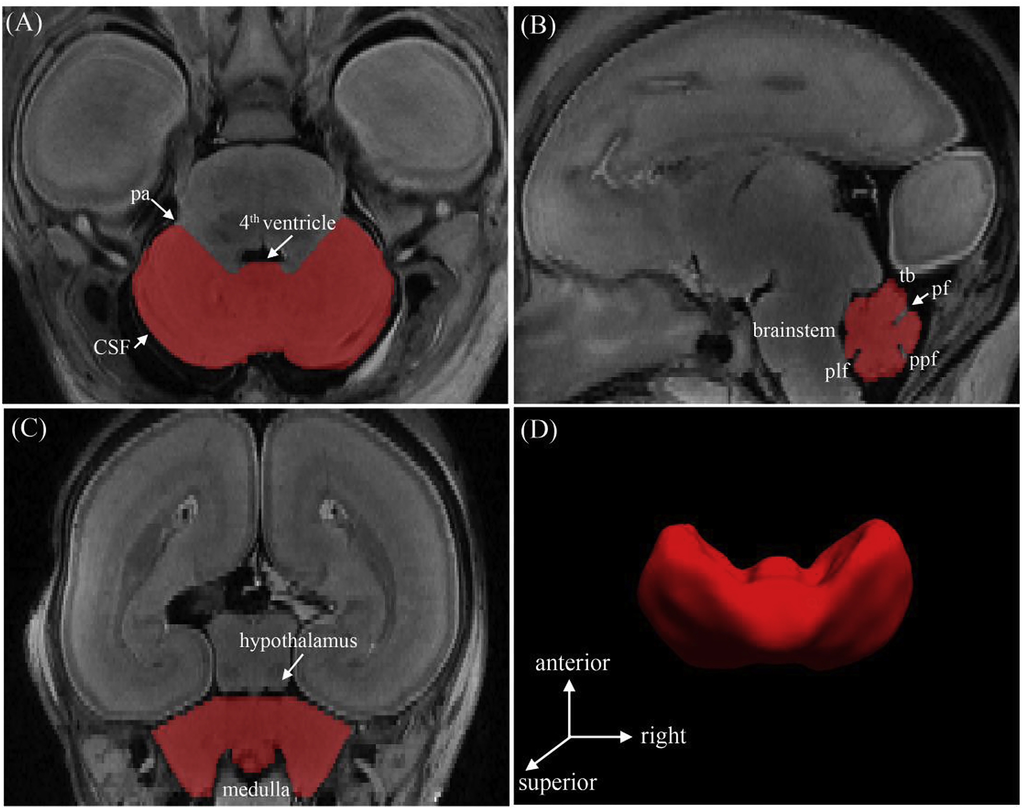
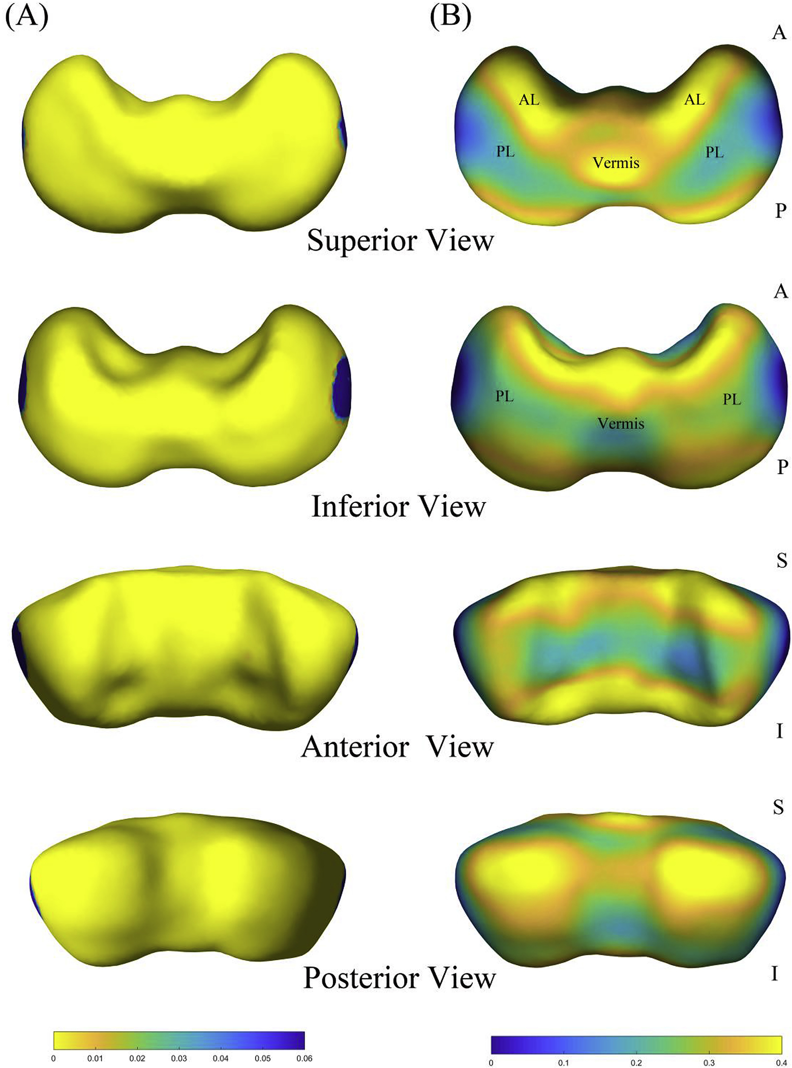
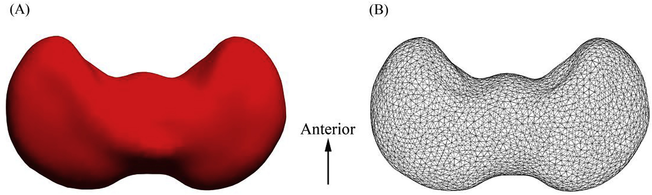
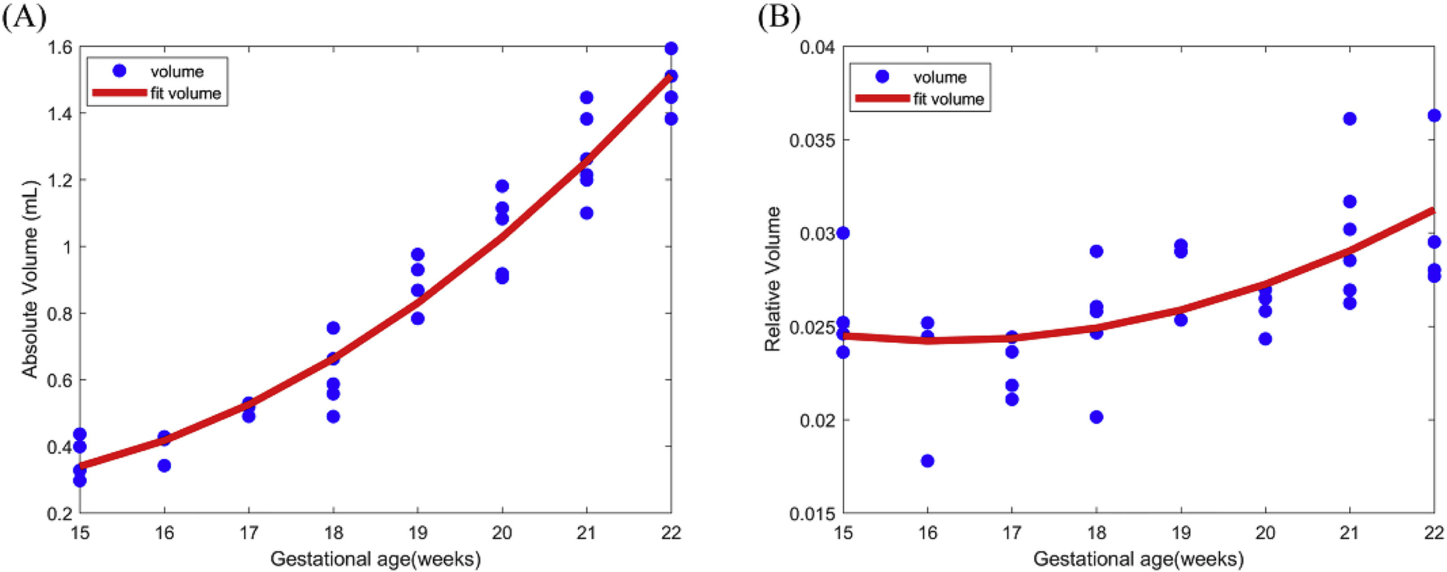
The protracted nature of development makes the cerebellum vulnerable to a broad spectrum of pathologic conditions, especially during the early fetal period. This study aims to characterize normal cerebellar growth in human fetuses during the early second trimester. We manually segmented the fetal cerebellum using 7.0-T high-resolution MR images obtained in 35 specimens with gestational ages ranging from 15 to 22 weeks. Volume measurements and shape analysis were performed to quantitatively evaluate global and regional cerebellar growth. The absolute volume of the fetal cerebellum showed a quadratic growth with increasing gestational age, while the pattern of relative volume changes revealed that the cerebellum grew at a greater pace than the cerebrum after 17 gestational weeks. Shape analysis was used to examine the distinctive development of subregions of the cerebellum. The extreme lateral portions of both cerebellar hemispheres showed the lowest rate of growth. The anterior lobe grew faster than most of the posterior lobe. These findings expand our understanding of the early growth pattern of the human cerebellum and could be further used to assess the developmental conditions of the fetal brain.

摘要

小脑的发育过程较为漫长,使其容易受到广泛的病理条件影响,尤其是在胎儿早期。本研究旨在描述人类胎儿在妊娠中期第二个 10 周时的正常小脑生长情况。我们使用 7.0-T 高分辨率磁共振成像对 35 个样本进行了手动分割,这些样本的胎龄范围从 15 周到 22 周。我们进行了体积测量和形状分析,以定量评估小脑的整体和区域生长。胎儿小脑的绝对体积随胎龄的增加呈二次生长,而相对体积变化的模式表明,小脑在 17 周后比大脑生长更快。形状分析用于检查小脑各亚区的独特发育。小脑两个半球的最外侧部分生长速度最慢。前叶比后叶的大部分区域生长更快。这些发现扩展了我们对人类小脑早期生长模式的理解,可进一步用于评估胎儿大脑的发育状况。

https://cdn.ncbi.nlm.nih.gov/pmc/blobs/daee/7055298/f7c46564675c/nihms-1064742-f0004.jpg
https://cdn.ncbi.nlm.nih.gov/pmc/blobs/daee/7055298/96a3d6c32d25/nihms-1064742-f0001.jpg
https://cdn.ncbi.nlm.nih.gov/pmc/blobs/daee/7055298/c6d2df85b858/nihms-1064742-f0002.jpg
https://cdn.ncbi.nlm.nih.gov/pmc/blobs/daee/7055298/e914dda75d71/nihms-1064742-f0003.jpg
https://cdn.ncbi.nlm.nih.gov/pmc/blobs/daee/7055298/f7c46564675c/nihms-1064742-f0004.jpg

相似文献

1
Morphometric development of the human fetal cerebellum during the early second trimester.人类胎儿小脑在第二个妊娠早期的形态发育。
Neuroimage. 2020 Feb 15;207:116372. doi: 10.1016/j.neuroimage.2019.116372. Epub 2019 Nov 18.
2
3D morphometric analysis of human fetal cerebellar development.三维形态计量分析人类胎儿小脑发育。
Cerebellum. 2012 Sep;11(3):761-70. doi: 10.1007/s12311-011-0338-2.
3
A magnetic resonance template for normal cerebellar development in the human fetus.人类胎儿正常小脑发育的磁共振模板。
Neurosurgery. 1997 Oct;41(4):924-8; discussion 928-9. doi: 10.1097/00006123-199710000-00029.
4
Fetal cerebellar volume and symmetry on 3-d ultrasound: volume measurement with multiplanar and vocal techniques.三维超声检查下胎儿小脑体积与对称性:多平面及容积技术测量体积
Ultrasound Med Biol. 2009 Aug;35(8):1284-9. doi: 10.1016/j.ultrasmedbio.2009.03.016. Epub 2009 Jun 21.
5
Association of gestational age with MRI-based biometrics of brain development in fetuses.胎儿脑发育的 MRI 生物测量与胎龄的关系。
BMC Med Imaging. 2020 Nov 25;20(1):125. doi: 10.1186/s12880-020-00525-9.
6
Development of the fetal cerebral cortex in the second trimester: assessment with 7T postmortem MR imaging.胎儿大脑皮质在妊娠中期的发育:7T 后尸体磁共振成像评估。
AJNR Am J Neuroradiol. 2013 Jul;34(7):1462-7. doi: 10.3174/ajnr.A3406. Epub 2013 Feb 14.
7
Quantitative MRI measurements of human fetal brain development in utero.子宫内人类胎儿脑发育的定量磁共振成像测量
Neuroimage. 2006 Nov 1;33(2):463-70. doi: 10.1016/j.neuroimage.2006.07.005. Epub 2006 Aug 30.
8
Altered local cerebellar and brainstem development in preterm infants.早产儿小脑和脑干局部发育改变。
Neuroimage. 2020 Jun;213:116702. doi: 10.1016/j.neuroimage.2020.116702. Epub 2020 Mar 5.
9
Fetal Brain Growth in the Early Second Trimester.胎儿大脑在孕早期的生长情况。
AJNR Am J Neuroradiol. 2023 Dec 11;44(12):1440-1444. doi: 10.3174/ajnr.A8051.
10
Development of the human fetal hippocampal formation during early second trimester.孕中期早期人类胎儿海马结构的发育
Neuroimage. 2015 Oct 1;119:33-43. doi: 10.1016/j.neuroimage.2015.06.055. Epub 2015 Jun 26.

引用本文的文献

1
The potential ameliorative effect of mesenchymal stem cells-derived exosomes on cerebellar histopathology and their modifying role on PI3k-mTOR signaling in rat model of autism spectrum disorder.间充质干细胞衍生外泌体对自闭症谱系障碍大鼠模型小脑组织病理学的潜在改善作用及其对PI3k-mTOR信号通路的调节作用。
J Mol Histol. 2025 Jan 6;56(1):65. doi: 10.1007/s10735-024-10335-w.
2
Structural deviations of the posterior fossa and the cerebellum and their cognitive links in a neurodevelopmental deletion syndrome.神经发育性缺失综合征中后颅窝和小脑的结构偏差及其与认知的关联。
Mol Psychiatry. 2024 Nov;29(11):3395-3411. doi: 10.1038/s41380-024-02584-8. Epub 2024 May 14.
3

本文引用的文献

1
Riemannian metric optimization on surfaces (RMOS) for intrinsic brain mapping in the Laplace-Beltrami embedding space.曲面的黎曼度量优化(RMOS)在拉普拉斯-贝尔特拉米嵌入空间中的内在脑图谱绘制。
Med Image Anal. 2018 May;46:189-201. doi: 10.1016/j.media.2018.03.004. Epub 2018 Mar 16.
2
Normative biometry of the fetal brain using magnetic resonance imaging.利用磁共振成像对胎儿大脑进行标准化生物测量
Brain Struct Funct. 2017 Jul;222(5):2295-2307. doi: 10.1007/s00429-016-1342-6. Epub 2016 Nov 24.
3
Prenatal Cerebellar Disruptions: Neuroimaging Spectrum of Findings in Correlation with Likely Mechanisms and Etiologies of Injury.
Morphological changes in the cerebellum during aging: evidence from convolutional neural networks and shape analysis.
衰老过程中小脑的形态学变化:来自卷积神经网络和形状分析的证据
Front Aging Neurosci. 2024 Apr 17;16:1359320. doi: 10.3389/fnagi.2024.1359320. eCollection 2024.
4
Fetal brain MRI atlases and datasets: A review.胎儿脑 MRI 图谱和数据集:综述。
Neuroimage. 2024 Apr 15;292:120603. doi: 10.1016/j.neuroimage.2024.120603. Epub 2024 Apr 6.
5
Preterm Birth Alters the Regional Development and Structural Covariance of Cerebellum at Term-Equivalent Age.早产儿在足月时其小脑的区域发育和结构协变发生改变。
Cerebellum. 2024 Oct;23(5):1932-1941. doi: 10.1007/s12311-024-01691-0. Epub 2024 Apr 6.
6
Aging pattern of the brainstem based on volumetric measurement and optimized surface shape analysis.基于容积测量和优化表面形态分析的脑干老化模式。
Brain Imaging Behav. 2024 Apr;18(2):396-411. doi: 10.1007/s11682-023-00840-z. Epub 2023 Dec 29.
7
Fetal Brain Growth in the Early Second Trimester.胎儿大脑在孕早期的生长情况。
AJNR Am J Neuroradiol. 2023 Dec 11;44(12):1440-1444. doi: 10.3174/ajnr.A8051.
8
Neuromorphological Atlas of Human Prenatal Brain Development: White Paper.《人类产前脑发育神经形态学图谱:白皮书》
Life (Basel). 2023 May 13;13(5):1182. doi: 10.3390/life13051182.
9
Assessment of longitudinal brain development using super-resolution magnetic resonance imaging following fetal surgery for open spina bifida.采用超分辨率磁共振成像评估开放性脊柱裂胎儿手术后的纵向脑发育。
Ultrasound Obstet Gynecol. 2023 Nov;62(5):707-720. doi: 10.1002/uog.26244.
10
Morphometry Difference of the Hippocampal Formation Between Blind and Sighted Individuals.盲人与视力正常者海马结构的形态测量差异
Front Neurosci. 2021 Nov 4;15:715749. doi: 10.3389/fnins.2021.715749. eCollection 2021.
产前小脑发育异常:与损伤可能机制及病因相关的神经影像学表现谱
Neuroimaging Clin N Am. 2016 Aug;26(3):359-72. doi: 10.1016/j.nic.2016.03.006.
4
Three-dimensional sonographic volume measurement of the fetal cerebellum.胎儿小脑的三维超声容积测量
J Med Ultrason (2001). 2007 Mar;34(1):17-21. doi: 10.1007/s10396-006-0122-y. Epub 2007 Mar 15.
5
Smaller Cerebellar Growth and Poorer Neurodevelopmental Outcomes in Very Preterm Infants Exposed to Neonatal Morphine.暴露于新生儿期吗啡的极早产儿小脑生长较小且神经发育结局较差。
J Pediatr. 2016 May;172:81-87.e2. doi: 10.1016/j.jpeds.2015.12.024. Epub 2016 Jan 4.
6
Cerebro-cerebellar circuits in autism spectrum disorder.自闭症谱系障碍中的大脑-小脑回路
Front Neurosci. 2015 Nov 5;9:408. doi: 10.3389/fnins.2015.00408. eCollection 2015.
7
Hindbrain regional growth in preterm newborns and its impairment in relation to brain injury.早产儿后脑区域的生长及其与脑损伤相关的损害。
Hum Brain Mapp. 2016 Feb;37(2):678-88. doi: 10.1002/hbm.23058. Epub 2015 Nov 21.
8
Consensus Paper: Cerebellar Development.共识文件:小脑发育
Cerebellum. 2016 Dec;15(6):789-828. doi: 10.1007/s12311-015-0724-2.
9
Development of the human fetal hippocampal formation during early second trimester.孕中期早期人类胎儿海马结构的发育
Neuroimage. 2015 Oct 1;119:33-43. doi: 10.1016/j.neuroimage.2015.06.055. Epub 2015 Jun 26.
10
The cerebellum, sensitive periods, and autism.小脑、敏感期与自闭症。
Neuron. 2014 Aug 6;83(3):518-32. doi: 10.1016/j.neuron.2014.07.016.