Döppler Heike, Bastea Ligia I, Borges Sahra, Spratley Samantha J, Pearce Sarah E, Storz Peter
Department of Cancer Biology, Mayo Clinic, Jacksonville, Florida, United States of America.
PLoS One. 2014 May 19;9(5):e98090. doi: 10.1371/journal.pone.0098090. eCollection 2014.
Protein kinase D (PKD) enzymes regulate cofilin-driven actin reorganization and directed cell migration through both p21-activated kinase 4 (PAK4) and the phosphatase slingshot 1L (SSH1L). The relative contributions of different endogenous PKD isoforms to both signaling pathways have not been elucidated, sufficiently.
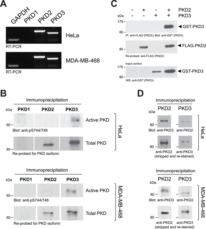
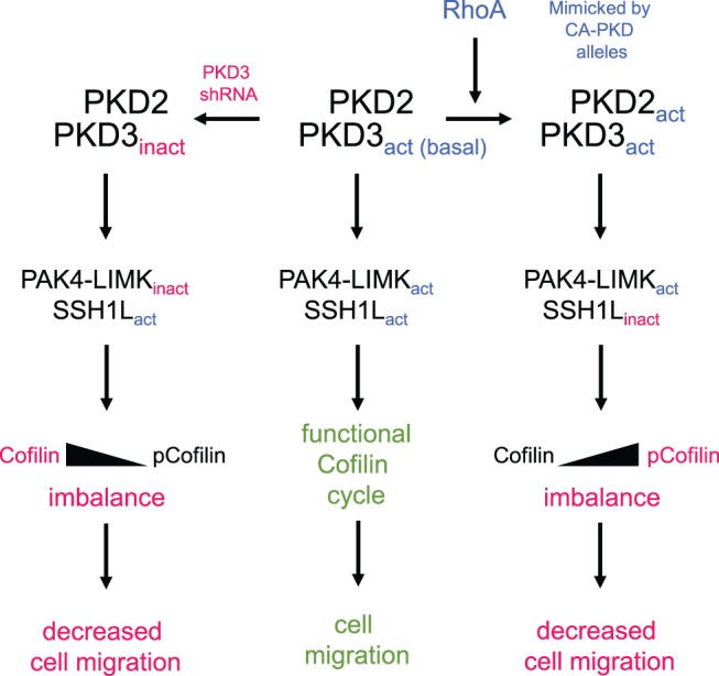
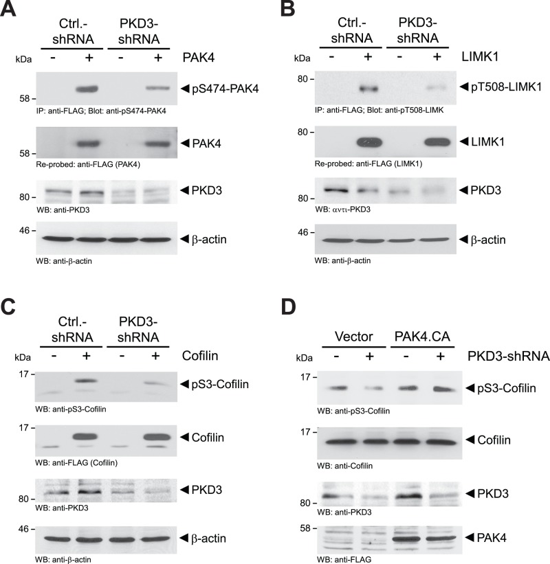
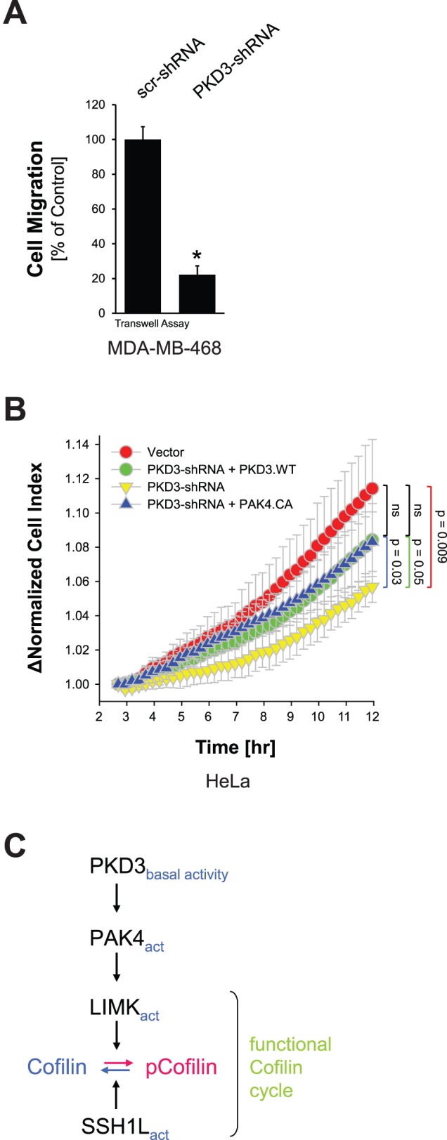
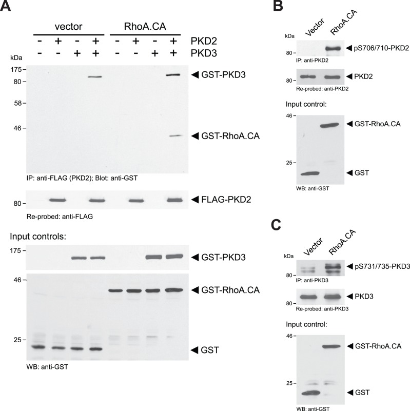
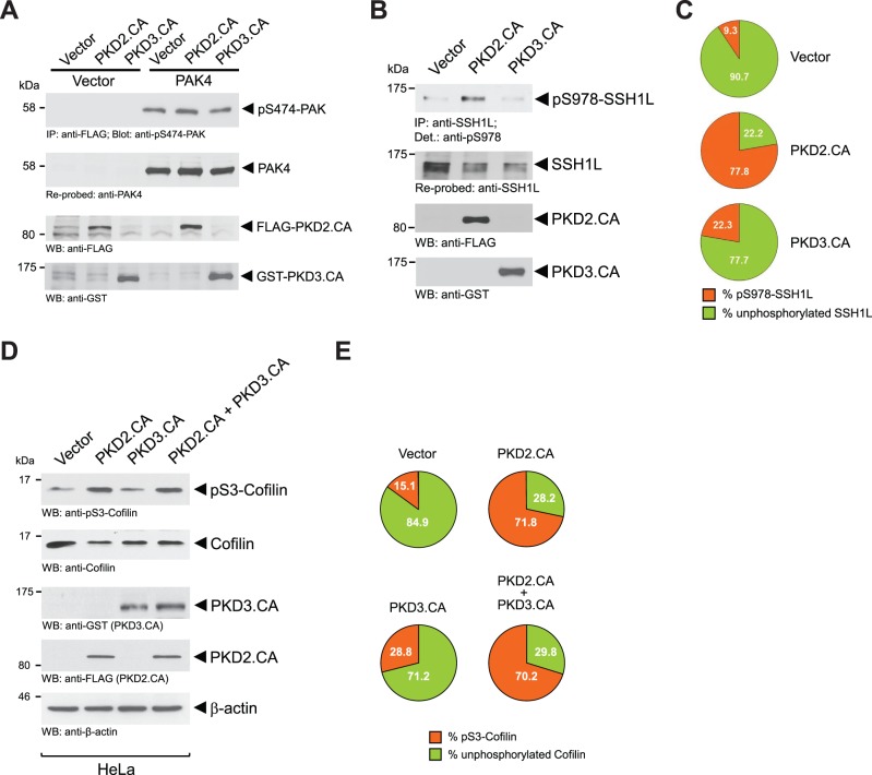
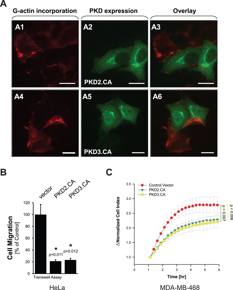
METHODOLOGY/PRINCIPAL FINDINGS: We here analyzed two cell lines (HeLa and MDA-MB-468) that express the subtypes protein kinase D2 (PKD2) and protein kinase D3 (PKD3). We show that under normal growth conditions both isoforms can form a complex, in which PKD3 is basally-active and PKD2 is inactive. Basal activity of PKD3 mediates PAK4 activity and downstream signaling, but does not significantly inhibit SSH1L. This signaling constellation was required for facilitating directed cell migration. Activation of PKD2 and further increase of PKD3 activity leads to additional phosphorylation and inhibition of endogenous SSH1L. Net effect is a dramatic increase in phospho-cofilin and a decrease in cell migration, since now both PAK4 and SSH1L are regulated by the active PKD2/PKD3 complex.
CONCLUSIONS/SIGNIFICANCE: Our data suggest that PKD complexes provide an interface for both cofilin regulatory pathways. Dependent on the activity of involved PKD enzymes signaling can be balanced to guarantee a functional cofilin activity cycle and increase cell migration, or imbalanced to decrease cell migration. Our data also provide an explanation of how PKD isoforms mediate different effects on directed cell migration.
蛋白激酶D(PKD)酶通过p21激活激酶4(PAK4)和磷酸酶弹弓1L(SSH1L)调节丝切蛋白驱动的肌动蛋白重组和定向细胞迁移。不同内源性PKD亚型对这两种信号通路的相对贡献尚未得到充分阐明。
方法/主要发现:我们在此分析了两种表达蛋白激酶D2(PKD2)和蛋白激酶D3(PKD3)亚型的细胞系(HeLa和MDA-MB-468)。我们发现,在正常生长条件下,这两种亚型均可形成复合物,其中PKD3具有基础活性,而PKD2无活性。PKD3的基础活性介导PAK4活性和下游信号传导,但对SSH1L无明显抑制作用。这种信号组合是促进定向细胞迁移所必需的。PKD2的激活以及PKD3活性的进一步增加会导致内源性SSH1L的额外磷酸化和抑制。最终效应是磷酸化丝切蛋白显著增加,细胞迁移减少,因为此时PAK4和SSH1L均受活性PKD2/PKD3复合物调控。
结论/意义:我们的数据表明,PKD复合物为丝切蛋白的两条调节通路提供了一个接口。根据所涉及的PKD酶的活性,信号传导可以保持平衡以确保丝切蛋白活性循环正常并促进细胞迁移,也可以失衡以减少细胞迁移。我们的数据还解释了PKD亚型如何介导对定向细胞迁移的不同影响。