Translational and Molecular Imaging Institute, Department of Radiology, Icahn School of Medicine at Mount Sinai, New York, New York, United States.
PLoS One. 2014 May 19;9(5):e97355. doi: 10.1371/journal.pone.0097355. eCollection 2014.
To quantify short-term reproducibility (in fasting conditions) and postprandial changes after a meal in portal vein (PV) flow parameters measured with phase contrast (PC) imaging, liver diffusion parameters measured with multiple b value diffusion-weighted imaging (DWI) and liver stiffness (LS) measured with MR elastography (MRE) in healthy volunteers and patients with liver disease at 3.0 T.
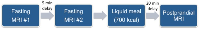
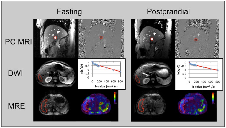
In this IRB-approved prospective study, 30 subjects (11 healthy volunteers and 19 liver disease patients; 23 males, 7 females; mean age 46.5 y) were enrolled. Imaging included 2D PC imaging, multiple b value DWI and MRE. Subjects were initially scanned twice in fasting state to assess short-term parameter reproducibility, and then scanned 20 min. after a liquid meal. PV flow/velocity, LS, liver true diffusion coefficient (D), pseudodiffusion coefficient (D*), perfusion fraction (PF) and apparent diffusion coefficient (ADC) were measured in fasting and postprandial conditions. Short-term reproducibility was assessed in fasting conditions by measuring coefficients of variation (CV) and Bland-Altman limits of agreement. Differences in MR metrics before and after caloric intake and between healthy volunteers and liver disease patients were assessed.
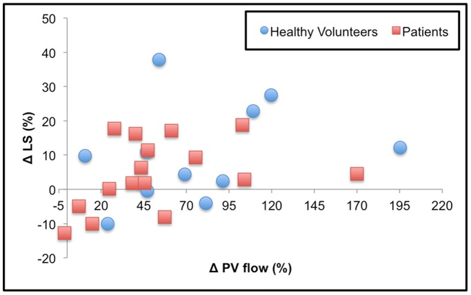
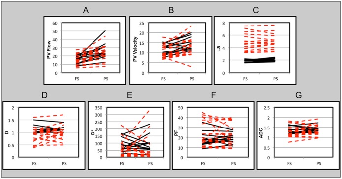
PV flow parameters, D, ADC and LS showed good to excellent short-term reproducibility in fasting state (CV <16%), while PF and D* showed acceptable and poor reproducibility (CV = 20.4% and 51.6%, respectively). PV flow parameters and LS were significantly higher (p<0.04) in postprandial state while liver diffusion parameters showed no significant change (p>0.2). LS was significantly higher in liver disease patients compared to healthy volunteers both in fasting and postprandial conditions (p<0.001). Changes in LS were significantly correlated with changes in PV flow (Spearman rho = 0.48, p = 0.013).
Caloric intake had no/minimal/large impact on diffusion/stiffness/portal vein flow, respectively. PC MRI and MRE but not DWI should be performed in controlled fasting state.
在 3.0T 磁共振扫描仪上,量化健康志愿者和肝病患者空腹(禁食状态)和餐后门静脉(PV)流量参数(使用相位对比(PC)成像测量)、肝脏扩散参数(使用多个 b 值扩散加权成像(DWI)测量)和肝脏硬度(LS)(使用磁共振弹性成像(MRE)测量)的短期重复性(在禁食状态下),以及餐后的变化。
本项经机构审查委员会批准的前瞻性研究共纳入 30 名受试者(11 名健康志愿者和 19 名肝病患者;23 名男性,7 名女性;平均年龄 46.5 岁)。成像包括二维 PC 成像、多 b 值 DWI 和 MRE。受试者首先在空腹状态下扫描两次,以评估短期参数的重复性,然后在液体餐后 20 分钟进行扫描。在空腹和餐后条件下测量 PV 流量/流速、LS、肝脏真实扩散系数(D)、假性扩散系数(D*)、灌注分数(PF)和表观扩散系数(ADC)。通过测量变异系数(CV)和 Bland-Altman 协议界限评估空腹状态下的短期重复性。评估热量摄入前后的 MR 指标差异以及健康志愿者和肝病患者之间的差异。
PV 流量参数、D、ADC 和 LS 在空腹状态下具有良好到极好的短期重复性(CV<16%),而 PF 和 D* 的重复性可接受和较差(CV=20.4%和 51.6%)。PV 流量参数和 LS 在餐后状态下显著升高(p<0.04),而肝脏扩散参数无明显变化(p>0.2)。LS 在空腹和餐后状态下均显著高于健康志愿者(p<0.001)。LS 的变化与 PV 流量的变化显著相关(Spearman rho=0.48,p=0.013)。
热量摄入对扩散/硬度/门静脉流量分别有最小/无/较大影响。PC MRI 和 MRE,但不是 DWI,应在受控的禁食状态下进行。