JARA - High Performance Computing, IT Center - Computational Science and Engineering, Computer Science Department, Virtual Reality Group, RWTH Aachen University Aachen, Germany.
JARA - Translational Brain Medicine, Institute of Neuroscience and Medicine (INM-1), Research Centre Jülich Jülich, Germany.
Front Neuroinform. 2014 May 5;8:42. doi: 10.3389/fninf.2014.00042. eCollection 2014.
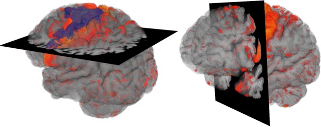
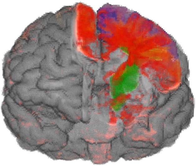
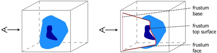
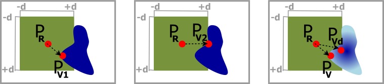
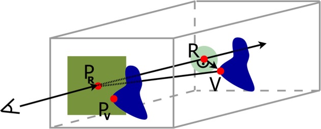
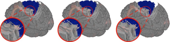
The visualization of the progression of brain tissue loss in neurodegenerative diseases like corticobasal syndrome (CBS) can provide not only information about the localization and distribution of the volume loss, but also helps to understand the course and the causes of this neurodegenerative disorder. The visualization of such medical imaging data is often based on 2D sections, because they show both internal and external structures in one image. Spatial information, however, is lost. 3D visualization of imaging data is capable to solve this problem, but it faces the difficulty that more internally located structures may be occluded by structures near the surface. Here, we present an application with two designs for the 3D visualization of the human brain to address these challenges. In the first design, brain anatomy is displayed semi-transparently; it is supplemented by an anatomical section and cortical areas for spatial orientation, and the volumetric data of volume loss. The second design is guided by the principle of importance-driven volume rendering: A direct line-of-sight to the relevant structures in the deeper parts of the brain is provided by cutting out a frustum-like piece of brain tissue. The application was developed to run in both, standard desktop environments and in immersive virtual reality environments with stereoscopic viewing for improving the depth perception. We conclude, that the presented application facilitates the perception of the extent of brain degeneration with respect to its localization and affected regions.
神经退行性疾病(如皮质基底节综合征,CBS)中脑组织损失进展的可视化不仅可以提供关于体积损失的定位和分布的信息,还有助于理解这种神经退行性疾病的过程和原因。这种医学成像数据的可视化通常基于 2D 切片,因为它们在一个图像中同时显示内部和外部结构。然而,空间信息会丢失。成像数据的 3D 可视化能够解决这个问题,但它面临着内部结构可能被表面附近结构遮挡的困难。在这里,我们提出了一个应用程序,有两种设计用于 3D 可视化人脑,以解决这些挑战。在第一种设计中,脑解剖结构以半透明方式显示;它通过解剖切片和皮质区域进行空间定位,以及体积损失的体积数据进行补充。第二种设计基于重要性驱动的体积渲染原理:通过切出一个类似截头锥体的脑组织块,为大脑深部的相关结构提供直接的视线。该应用程序旨在运行在标准桌面环境和具有立体观看的沉浸式虚拟现实环境中,以提高深度感知能力。我们得出结论,所提出的应用程序有助于感知大脑退化的程度及其定位和受影响的区域。