Brand Christian, Abdel-Atti Dalya, Zhang Yachao, Carlin Sean, Clardy Susan M, Keliher Edmund J, Weber Wolfgang A, Lewis Jason S, Reiner Thomas
Radiochemistry and Imaging Sciences Service and §Molecular Imaging and Therapy Service, Department of Radiology, ∥Molecular Pharmacology and Chemistry Program, and ⊥Center for Molecular Imaging and Nanotechnology, Memorial Sloan Kettering Cancer Center , New York, New York 10065, United States.
Bioconjug Chem. 2014 Jul 16;25(7):1323-30. doi: 10.1021/bc500178d. Epub 2014 Jun 13.
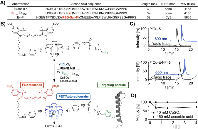
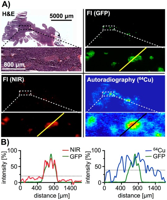
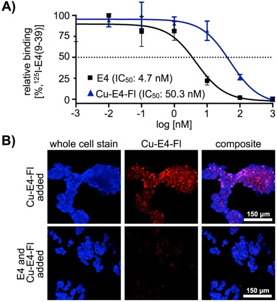
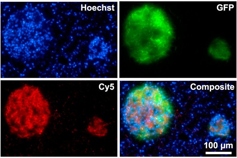
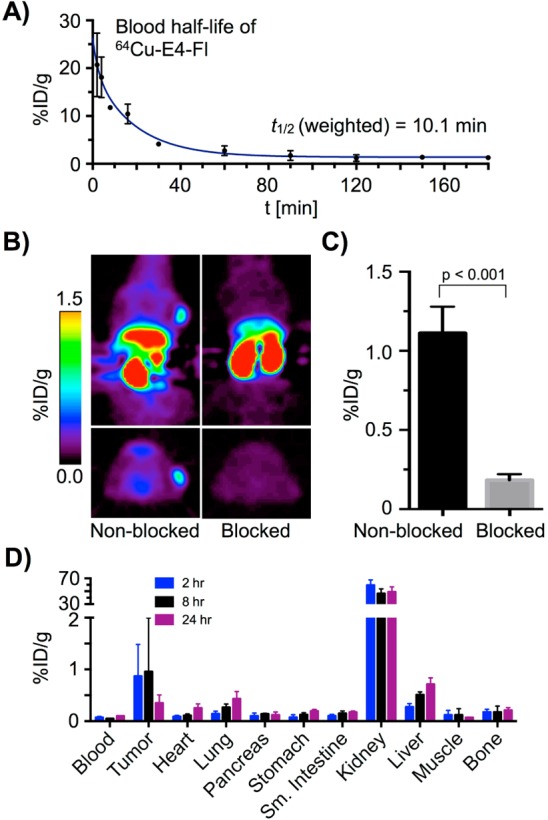
Accurate visualization and quantification of β-cell mass is critical for the improved understanding, diagnosis, and treatment of both type 1 diabetes (T1D) and insulinoma. Here, we describe the synthesis of a bimodal imaging probe (PET/fluorescence) for imaging GLP-1R expression in the pancreas and in pancreatic islet cell tumors. The conjugation of a bimodal imaging tag containing a near-infrared fluorescent dye, and the copper chelator sarcophagine to the GLP-1R targeting peptide exendin-4 provided the basis for the bimodal imaging probe. Conjugation was performed via a novel sequential one-pot synthetic procedure including (64)Cu radiolabeling and copper-catalyzed click-conjugation. The bimodal imaging agent (64)Cu-E4-Fl was synthesized in good radiochemical yield and specific activity (RCY = 36%, specific activity: 141 μCi/μg, >98% radiochemical purity). The agent showed good performance in vivo and ex vivo, visualizing small xenografts (<2 mm) with PET and pancreatic β-cell mass by phosphor autoradiography. Using the fluorescent properties of the probe, we were able to detect individual pancreatic islets, confirming specific binding to GLP-1R and surpassing the sensitivity of the radioactive label. The use of bimodal PET/fluorescent imaging probes is promising for preoperative imaging and fluorescence-assisted analysis of patient tissues. We believe that our procedure could become relevant as a protocol for the development of bimodal imaging agents.
准确可视化和定量β细胞质量对于更好地理解、诊断和治疗1型糖尿病(T1D)和胰岛素瘤至关重要。在此,我们描述了一种用于胰腺和胰岛细胞瘤中GLP-1R表达成像的双模态成像探针(PET/荧光)的合成。将含有近红外荧光染料的双模态成像标签和铜螯合剂肌氨酸与GLP-1R靶向肽艾塞那肽-4偶联,为双模态成像探针提供了基础。通过一种新颖的连续一锅法合成程序进行偶联,包括(64)Cu放射性标记和铜催化的点击偶联。双模态成像剂(64)Cu-E4-Fl以良好的放射化学产率和比活度合成(RCY = 36%,比活度:141 μCi/μg,放射化学纯度>98%)。该试剂在体内和体外均表现出良好的性能,通过PET可视化小的异种移植瘤(<2 mm),并通过磷光放射自显影术可视化胰腺β细胞质量。利用探针的荧光特性,我们能够检测单个胰岛,证实其与GLP-1R的特异性结合,并超过了放射性标记的灵敏度。双模态PET/荧光成像探针在术前成像和患者组织的荧光辅助分析方面具有广阔前景。我们相信,我们的方法可能成为双模态成像剂开发方案的相关方法。