Kim Ji Eun, Lee Sang Mok, Kim Soo Hyun, Tatman Phil, Gee Albert O, Kim Deok-Ho, Lee Kyung Eun, Jung Youngmee, Kim Sang Jun
Biomaterials Research Center, Korea Institute of Science and Technology, Seoul, South Korea.
Department of Physical and Rehabilitation Medicine, Samsung Medical Center, Seoul, South Korea.
Int J Nanomedicine. 2014 May 7;9 Suppl 1(Suppl 1):141-57. doi: 10.2147/IJN.S54114. eCollection 2014.
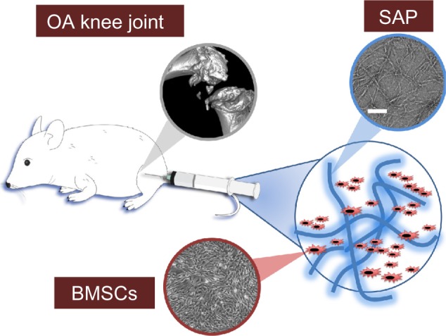
To evaluate the efficacy of mesenchymal stem cells (MSCs) encapsulated in self-assembled peptide (SAP) hydrogels in a rat knee model for the prevention of osteoarthritis (OA) progression.
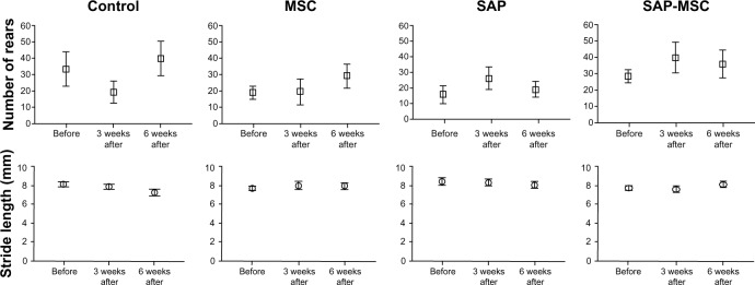
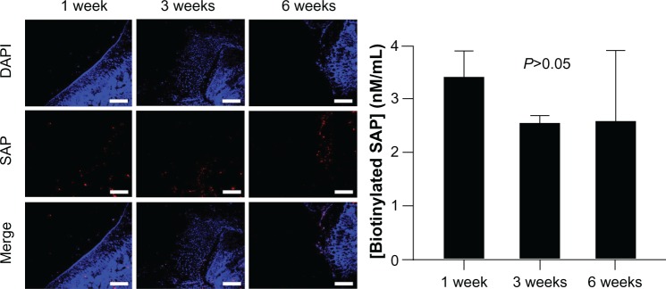
Nanostructured KLD-12 SAPs were used as the injectable hydrogels. Thirty-three Sprague Dawley rats were used for the OA model. Ten rats were used for the evaluation of biotin-tagged SAP disappearance. Twenty-three rats were divided into four groups: MSC (n=6), SAP (n=6), SAP-MSC (n=6), and no treatment (n=5). MSCs, SAPs, and SAP-MSCs were injected into the knee joints 3 weeks postsurgery. Histologic examination, immunofluorescent staining, measurement of cytokine levels, and micro-computed tomography analysis were conducted 6 weeks after injections. Behavioral studies were done to establish baseline measurements before treatment, and repeated 3 and 6 weeks after treatment to measure the efficacy of SAP-MSCs.
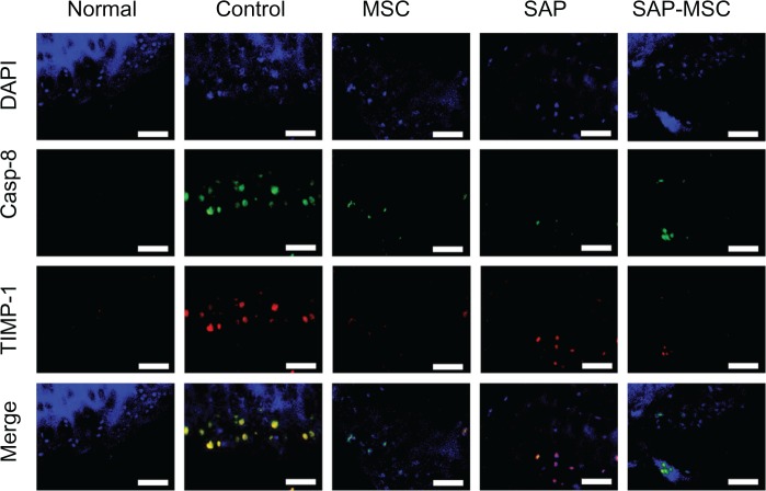
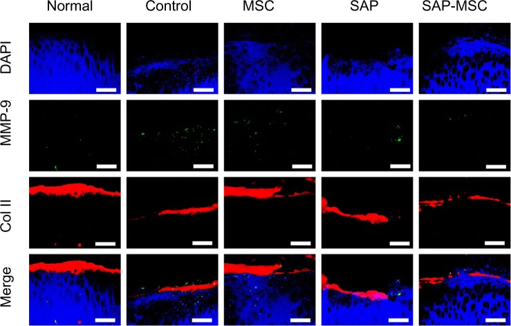
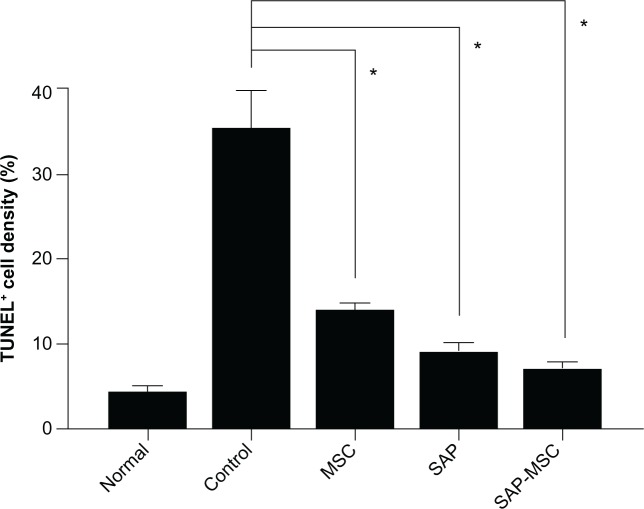
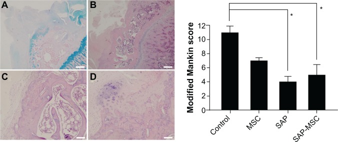
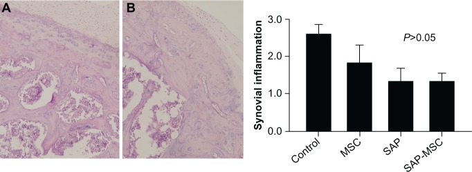
Concentration of biotinylated SAP at week 1 was not significantly different from those at week 3 and week 6 (P=0.565). Bone mineral density was significantly lower in SAP-MSC groups than controls (P=0.002). Significant differences in terminal deoxynucleotidyl transferase deoxyuridine triphosphate nick-end labeling staining between the control group and all other groups were observed. Caspase-8, tissue inhibitor of metalloproteinases 1, and matrix metalloproteinase 9 were diffusely stained in controls, whereas localized or minimal staining was observed in other groups. Modified Mankin scores were significantly lower in the SAP and SAP-MSC groups than in controls (P=0.001 and 0.013). Although not statistically significant, synovial inflammation scores were lower in the SAP (1.3±0.3) and SAP-MSC (1.3±0.2) groups than in controls (2.6±0.2). However, neither the cytokine level nor the behavioral score was significantly different between groups.
Injection of SAP-MSC hydrogels showed evidence of chondroprotection, as measured by the histologic grading and decreased expression of biochemical markers of inflammation and apoptosis. It also lowered subchondral bone mineral density, which can be increased by OA. This suggests that the SAP-MSC complex may have clinical potential to inhibit OA progression.
在大鼠膝关节模型中评估包裹于自组装肽(SAP)水凝胶中的间充质干细胞(MSC)预防骨关节炎(OA)进展的疗效。
纳米结构的KLD-12 SAP用作可注射水凝胶。33只Sprague Dawley大鼠用于OA模型。10只大鼠用于评估生物素标记的SAP消失情况。23只大鼠分为四组:MSC组(n = 6)、SAP组(n = 6)、SAP-MSC组(n = 6)和未治疗组(n = 5)。术后3周将MSC、SAP和SAP-MSC注射到膝关节中。注射后6周进行组织学检查、免疫荧光染色、细胞因子水平测量和微计算机断层扫描分析。在治疗前进行行为学研究以建立基线测量值,并在治疗后3周和6周重复进行以测量SAP-MSC的疗效。
第1周时生物素化SAP的浓度与第3周和第6周时无显著差异(P = 0.565)。SAP-MSC组的骨矿物质密度显著低于对照组(P = 0.002)。观察到对照组与所有其他组之间在末端脱氧核苷酸转移酶脱氧尿苷三磷酸缺口末端标记染色上存在显著差异。对照组中Caspase-8、金属蛋白酶组织抑制剂1和基质金属蛋白酶9呈弥漫性染色,而其他组观察到局部或最小程度的染色。SAP组和SAP-MSC组的改良Mankin评分显著低于对照组(P = 0.001和0.013)。虽然无统计学意义,但SAP组(1.3±0.3)和SAP-MSC组(1.3±0.2)的滑膜炎症评分低于对照组(2.6±0.2)。然而,各组之间细胞因子水平和行为评分均无显著差异。
通过组织学分级以及炎症和凋亡生化标志物表达降低来衡量,注射SAP-MSC水凝胶显示出软骨保护的证据。它还降低了软骨下骨矿物质密度,而OA可使其升高。这表明SAP-MSC复合物可能具有抑制OA进展的临床潜力。