Aerts Hugo J W L, Velazquez Emmanuel Rios, Leijenaar Ralph T H, Parmar Chintan, Grossmann Patrick, Carvalho Sara, Bussink Johan, Monshouwer René, Haibe-Kains Benjamin, Rietveld Derek, Hoebers Frank, Rietbergen Michelle M, Leemans C René, Dekker Andre, Quackenbush John, Gillies Robert J, Lambin Philippe
1] Department of Radiation Oncology (MAASTRO), Research Institute GROW, Maastricht University, 6229ET Maastricht, The Netherlands [2] Department of Radiation Oncology, Dana-Farber Cancer Institute, Brigham and Women's Hospital, Harvard Medical School, Boston, Massachusetts 02215-5450, USA [3] Department of Radiology, Dana-Farber Cancer Institute, Brigham and Women's Hospital, Harvard Medical School, Boston, Massachusetts 02215-5450, USA [4] Department of Biostatistics & Computational Biology, Dana-Farber Cancer Institute, Boston, Massachusetts 02215-5450, USA [5].
1] Department of Radiation Oncology (MAASTRO), Research Institute GROW, Maastricht University, 6229ET Maastricht, The Netherlands [2] Department of Radiation Oncology, Dana-Farber Cancer Institute, Brigham and Women's Hospital, Harvard Medical School, Boston, Massachusetts 02215-5450, USA [3].
Nat Commun. 2014 Jun 3;5:4006. doi: 10.1038/ncomms5006.
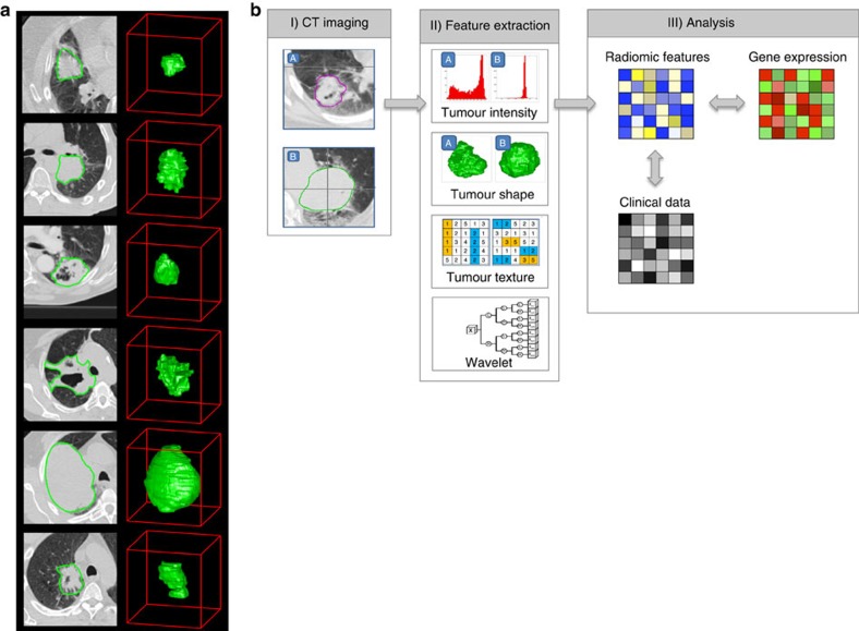
Human cancers exhibit strong phenotypic differences that can be visualized noninvasively by medical imaging. Radiomics refers to the comprehensive quantification of tumour phenotypes by applying a large number of quantitative image features. Here we present a radiomic analysis of 440 features quantifying tumour image intensity, shape and texture, which are extracted from computed tomography data of 1,019 patients with lung or head-and-neck cancer. We find that a large number of radiomic features have prognostic power in independent data sets of lung and head-and-neck cancer patients, many of which were not identified as significant before. Radiogenomics analysis reveals that a prognostic radiomic signature, capturing intratumour heterogeneity, is associated with underlying gene-expression patterns. These data suggest that radiomics identifies a general prognostic phenotype existing in both lung and head-and-neck cancer. This may have a clinical impact as imaging is routinely used in clinical practice, providing an unprecedented opportunity to improve decision-support in cancer treatment at low cost.
人类癌症表现出明显的表型差异,可通过医学成像进行无创可视化。放射组学是指通过应用大量定量图像特征对肿瘤表型进行全面量化。在此,我们对440个量化肿瘤图像强度、形状和纹理的特征进行了放射组学分析,这些特征是从1019例肺癌或头颈癌患者的计算机断层扫描数据中提取的。我们发现,大量放射组学特征在肺癌和头颈癌患者的独立数据集中具有预后价值,其中许多特征以前未被确定为具有显著性。放射基因组学分析表明,一个捕捉肿瘤内异质性的预后放射组学特征与潜在的基因表达模式相关。这些数据表明,放射组学识别出一种在肺癌和头颈癌中都存在的一般预后表型。这可能具有临床意义,因为成像在临床实践中经常使用,为以低成本改善癌症治疗中的决策支持提供了前所未有的机会。