Institute of Neuroscience and Medicine - 4, Forschungszentrum Jülich GmbH Jülich, Germany ; Department of Psychiatry, Psychotherapy and Psychosomatics, RWTH Aachen University Aachen, Germany ; JARA - Translational Brain Medicine Aachen, Germany.
Institute of Neuroscience and Medicine - 4, Forschungszentrum Jülich GmbH Jülich, Germany ; Department of Neurology, RWTH Aachen University Aachen, Germany.
Front Hum Neurosci. 2014 May 28;8:362. doi: 10.3389/fnhum.2014.00362. eCollection 2014.
Tourette syndrome (TS) is a neuropsychiatric disorder with the core phenomenon of tics, whose origin and temporal pattern are unclear. We investigated the When and Where of tic generation and resting state networks (RSNs) via functional magnetic resonance imaging (fMRI).
Tic-related activity and the underlying RSNs in adult TS were studied within one fMRI session. Participants were instructed to lie in the scanner and to let tics occur freely. Tic onset times, as determined by video-observance were used as regressors and added to preceding time-bins of 1 s duration each to detect prior activation. RSN were identified by independent component analysis (ICA) and correlated to disease severity by the means of dual regression.
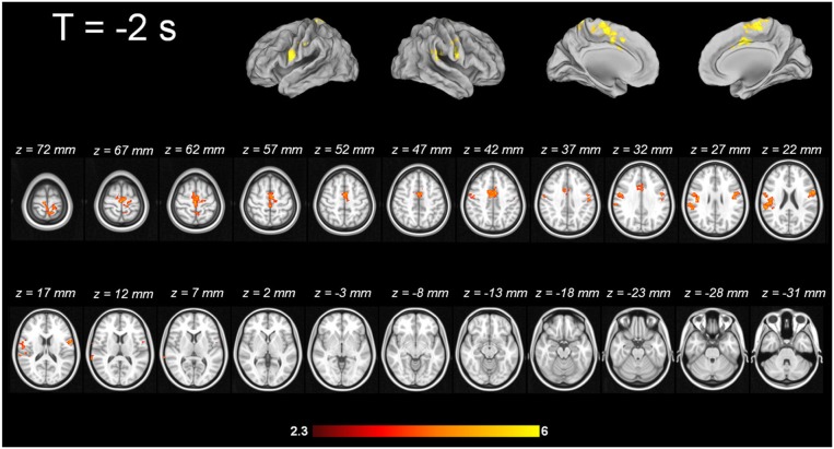
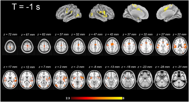
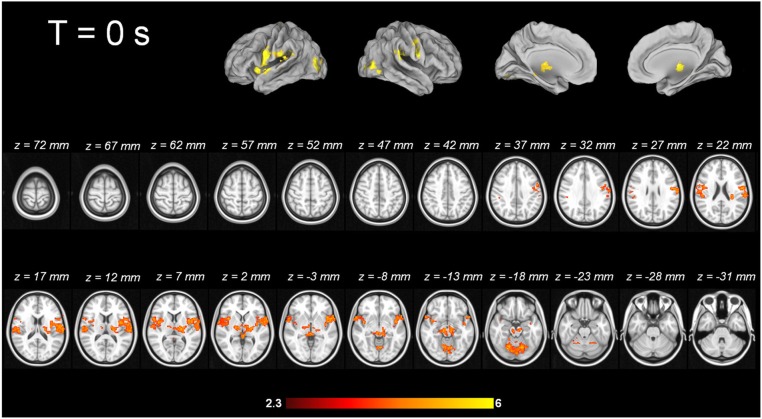
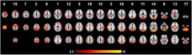
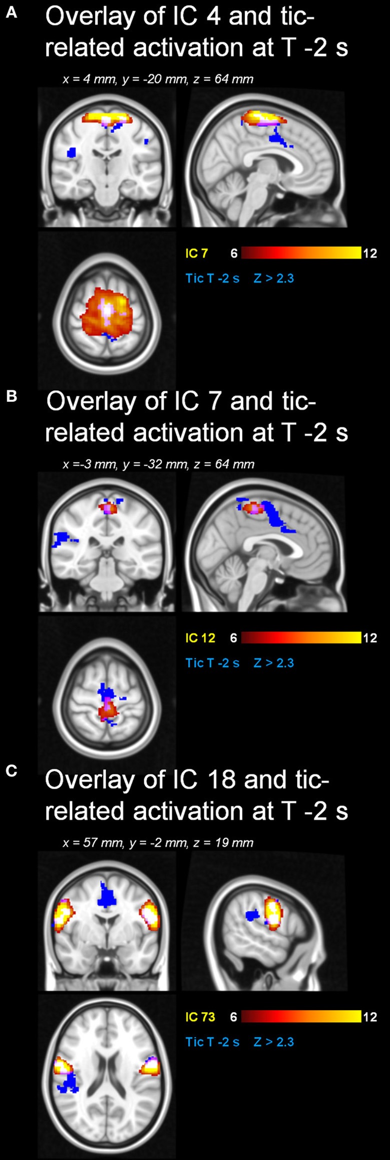
Two seconds before a tic, the supplementary motor area (SMA), ventral primary motor cortex, primary sensorimotor cortex and parietal operculum exhibited activation; 1 s before a tic, the anterior cingulate, putamen, insula, amygdala, cerebellum and the extrastriatal-visual cortex exhibited activation; with tic-onset, the thalamus, central operculum, primary motor and somatosensory cortices exhibited activation. Analysis of resting state data resulted in 21 components including the so-called default-mode network. Network strength in those regions in SMA of two premotor ICA maps that were also active prior to tic occurrence, correlated significantly with disease severity according to the Yale Global Tic Severity Scale (YGTTS) scores.
We demonstrate that the temporal pattern of tic generation follows the cortico-striato-thalamo-cortical circuit, and that cortical structures precede subcortical activation. The analysis of spontaneous fluctuations highlights the role of cortical premotor structures. Our study corroborates the notion of TS as a network disorder in which abnormal RSN activity might contribute to the generation of tics in SMA.
妥瑞氏综合征(TS)是一种神经精神疾病,其核心现象是抽搐,其起源和时间模式尚不清楚。我们通过功能磁共振成像(fMRI)研究了抽搐发作的时间和空间以及静息状态网络(RSN)。
在一次 fMRI 扫描中研究了成年 TS 患者的抽搐相关活动及其潜在的 RSN。参与者被指示躺在扫描仪中,让抽搐自由发生。通过视频观察确定抽搐发作时间,并将其用作回归器,然后将每个 1 秒的时间窗添加到每个时间窗中,以检测先前的激活。通过独立成分分析(ICA)确定 RSN,并通过双回归的方法将其与疾病严重程度相关联。
在抽搐前 2 秒,辅助运动区(SMA)、腹侧初级运动皮层、初级感觉运动皮层和顶叶岛盖区显示出激活;在抽搐前 1 秒,前扣带、壳核、岛叶、杏仁核、小脑和额外视觉皮层显示出激活;抽搐发作时,丘脑、中央岛盖、初级运动和体感皮层显示出激活。对静息状态数据的分析产生了 21 个成分,包括所谓的默认模式网络。根据耶鲁总体抽搐严重程度量表(YGTTS)评分,在与 tic 发生前同样活跃的两个前运动 ICA 图谱的 SMA 中,这些区域的网络强度与疾病严重程度显著相关。
我们证明了 tic 发作的时间模式遵循皮质-纹状体-丘脑-皮质回路,并且皮质结构先于皮质下激活。自发性波动的分析强调了皮质前运动结构的作用。我们的研究支持了 TS 作为一种网络障碍的观点,即异常的 RSN 活动可能有助于 SMA 中 tic 的产生。