Sackler Centre for Consciousness Science, University of Sussex, Falmer BN1 9RY, UK.
Department of Neuroscience, Brighton & Sussex Medical School, Falmer BN1 9RY, UK.
Neuroimage Clin. 2017 Aug 3;16:257-267. doi: 10.1016/j.nicl.2017.08.004. eCollection 2017.
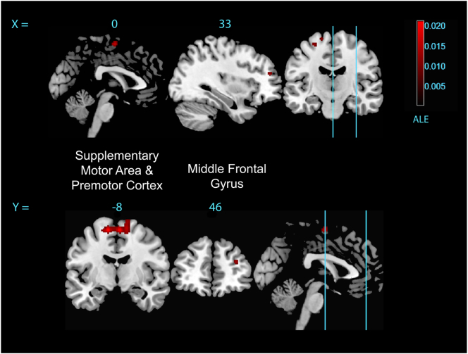
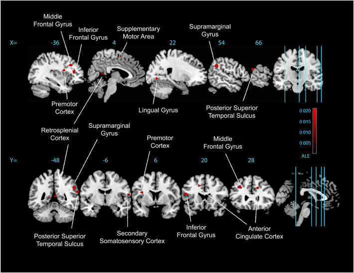
Tourette Syndrome (TS) is a neurodevelopmental condition characterized by chronic multiple tics, which are experienced as compulsive and 'unwilled'. Patients with TS can differ markedly in the frequency, severity, and bodily distribution of tics. Moreover, there are high comorbidity rates with attention deficit hyperactivity disorder (ADHD), obsessive compulsive disorder (OCD), anxiety disorders, and depression. This complex clinical profile may account for apparent variability of findings across neuroimaging studies that connect neural function to cognitive and motor behavior in TS. Here we crystalized information from neuroimaging regarding the functional circuitry of TS, and furthermore, tested specifically for neural determinants of tic severity, by applying activation likelihood estimation (ALE) meta-analyses to neuroimaging (activation) studies of TS. Fourteen task-based studies (13 fMRI and one H2O-PET) met rigorous inclusion criteria. These studies, encompassing 25 experiments and 651 participants, tested for differences between TS participants and healthy controls across cognitive, motor, perceptual and somatosensory domains. Relative to controls, TS participants showed distributed differences in the activation of prefrontal (inferior, middle, and superior frontal gyri), anterior cingulate, and motor preparation cortices (lateral premotor cortex and supplementary motor area; SMA). Differences also extended into sensory (somatosensory cortex and the lingual gyrus; V4); and temporo-parietal association cortices (posterior superior temporal sulcus, supramarginal gyrus, and retrosplenial cortex). Within TS participants, tic severity (reported using the Yale Global Tic Severity Scale; YGTSS) selectively correlated with engagement of SMA, precentral gyrus, and middle frontal gyrus across tasks. The dispersed involvement of multiple cortical regions with differences in functional reactivity may account for heterogeneity in the symptomatic expression of TS and its comorbidities. More specifically for tics and tic severity, the findings reinforce previously proposed contributions of premotor and lateral prefrontal cortices to tic expression.
妥瑞氏症(TS)是一种神经发育疾病,其特征为慢性多发性抽搐,这些抽搐被体验为强迫性和“不由自主”的。患有 TS 的患者在抽搐的频率、严重程度和身体分布上可能有很大的差异。此外,TS 还与注意力缺陷多动障碍(ADHD)、强迫症(OCD)、焦虑症和抑郁症的发病率很高。这种复杂的临床特征可能解释了神经影像学研究中连接神经功能与 TS 认知和运动行为的发现存在明显的可变性。在这里,我们总结了神经影像学关于 TS 功能回路的信息,并通过对 TS 的神经影像学(激活)研究应用激活似然估计(ALE)元分析,专门测试了抽动严重程度的神经决定因素。14 项基于任务的研究(13 项 fMRI 和 1 项 H2O-PET)符合严格的纳入标准。这些研究涵盖了 25 项实验和 651 名参与者,测试了 TS 参与者与健康对照组在认知、运动、感知和躯体感觉领域的差异。与对照组相比,TS 参与者在额前皮质(额下回、额中回和额上回)、前扣带皮质和运动准备皮质(外侧运动前皮质和补充运动区;SMA)的激活方面表现出分布上的差异。差异还扩展到感觉(躯体感觉皮质和舌回;V4)和颞顶联合皮质(后上颞回、缘上回和后扣带回)。在 TS 参与者中,抽动严重程度(使用耶鲁总体抽动严重程度量表报告;YGTSS)与跨任务 SMA、中央前回和额中回的参与选择性相关。多个皮质区域的功能反应差异的分散参与可能解释了 TS 及其共病的症状表达的异质性。更具体地说,对于抽动和抽动严重程度,这些发现强化了之前提出的运动前皮质和外侧前额叶皮质对抽动表达的贡献。