University of Queensland Centre for Clinical Research, Centre for Clinical Diagnostics, Royal Brisbane and Women's Hospital, Queensland, Australia.
Department of Obstetric and Gynaecology, Faculty of Medicine, Universidad de los Andes, Santiago, Chile.
PLoS One. 2014 Jun 6;9(6):e98667. doi: 10.1371/journal.pone.0098667. eCollection 2014.
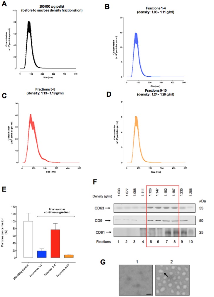
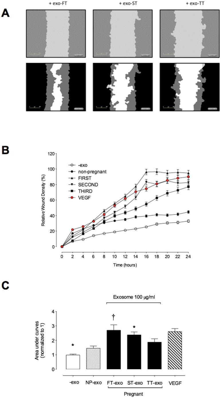
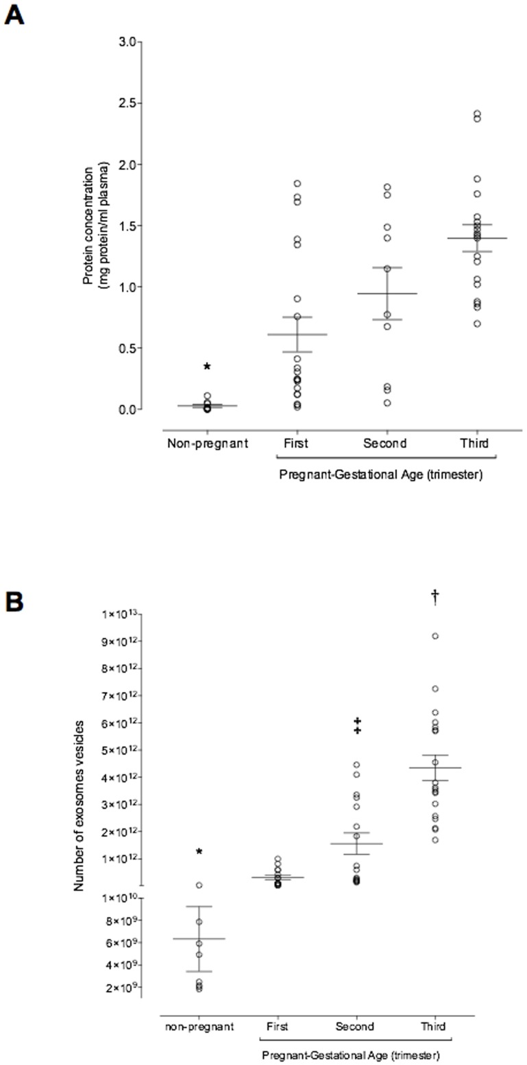
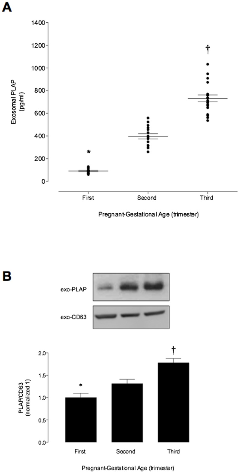
Studies completed to date provide persuasive evidence that placental cell-derived exosomes play a significant role in intercellular communication pathways that potentially contribute to placentation and development of materno-fetal vascular circulation. The aim of this study was to establish the gestational-age release profile and bioactivity of placental cell-derived exosome in maternal plasma. Plasma samples (n = 20 per pregnant group) were obtained from non-pregnant and pregnant women in the first (FT, 6-12 weeks), second (ST, 22-24 weeks) and third (TT, 32-38 weeks) trimester. The number of exosomes and placental exosome contribution were determined by quantifying immunoreactive exosomal CD63 and placenta-specific marker (PLAP), respectively. The effect of exosomes isolated from FT, ST and TT on endothelial cell migration were established using a real-time, live-cell imaging system (Incucyte). Exosome plasma concentration was more than 50-fold greater in pregnant women than in non-pregnant women (p<0.001). During normal healthy pregnancy, the number of exosomes present in maternal plasma increased significantly with gestational age by more that two-fold (p<0.001). Exosomes isolated from FT, ST and TT increased endothelial cell migration by 1.9±0.1, 1.6±0.2 and 1.3±0.1-fold, respectively compared to the control. Pregnancy is associated with a dramatic increase in the number of exosomes present in plasma and maternal plasma exosomes are bioactive. While the role of placental cell-derived exosome in regulating maternal and/or fetal vascular responses remains to be elucidated, changes in exosome profile may be of clinical utility in the diagnosis of placental dysfunction.
迄今为止完成的研究提供了有说服力的证据,证明胎盘细胞衍生的外泌体在细胞间通讯途径中发挥重要作用,这些途径可能有助于胎盘形成和母胎血管循环的发育。本研究旨在确定母体外周血浆中胎盘细胞衍生外泌体的妊娠龄释放特征和生物活性。从非妊娠和妊娠妇女(FT,6-12 周;ST,22-24 周;TT,32-38 周)获得血浆样本(每组 20 例)。通过分别定量免疫反应性外泌体 CD63 和胎盘特异性标志物(PLAP)来确定外泌体的数量和胎盘外泌体的贡献。使用实时活细胞成像系统(Incucyte)确定从 FT、ST 和 TT 分离的外泌体对内皮细胞迁移的影响。外泌体在血浆中的浓度在孕妇中比非孕妇高 50 多倍(p<0.001)。在正常健康妊娠期间,母体外周血浆中外泌体的数量随着妊娠龄的增加而显著增加,增加了两倍以上(p<0.001)。与对照组相比,FT、ST 和 TT 分离的外泌体分别使内皮细胞迁移增加 1.9±0.1、1.6±0.2 和 1.3±0.1 倍。妊娠与血浆中外泌体数量的急剧增加相关,并且母体外周血浆外泌体具有生物活性。虽然胎盘细胞衍生的外泌体在调节母体和/或胎儿血管反应中的作用仍有待阐明,但外泌体谱的变化可能对胎盘功能障碍的诊断具有临床应用价值。