Brown Matthew L, Yukata Kiminori, Farnsworth Christopher W, Chen Ding-Geng, Awad Hani, Hilton Matthew J, O'Keefe Regis J, Xing Lianping, Mooney Robert A, Zuscik Michael J
Center for Musculoskeletal Research, University of Rochester Medical Center, Rochester, New York, United States of America; School of Medicine and Dentistry, University of Rochester Medical Center, Rochester, New York, United States of America.
Center for Musculoskeletal Research, University of Rochester Medical Center, Rochester, New York, United States of America.
PLoS One. 2014 Jun 9;9(6):e99656. doi: 10.1371/journal.pone.0099656. eCollection 2014.
Impaired healing and non-union of skeletal fractures is a major public health problem, with morbidity exacerbated in patients with diabetes mellitus (DM). DM is prevalent worldwide and affects approximately 25.8 million US adults, with >90% having obesity-related type 2 DM (T2DM). While fracture healing in type 1 DM (T1DM) has been studied using animal models, an investigation into delayed healing in an animal model of T2DM has not yet been performed.
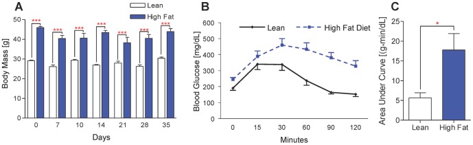
Male C57BL/6J mice at 5 weeks of age were placed on either a control lean diet or an experimental high-fat diet (HFD) for 12 weeks. A mid-diaphyseal open tibia fracture was induced at 17 weeks of age and a spinal needle was used for intra-medullary fixation. Mice were sacrificed at days 7, 10, 14, 21, 28, and 35 for micro-computed tomography (μCT), histology-based histomorphometry and molecular analyses, and biomechanical testing.
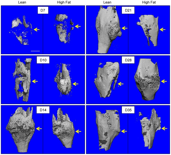
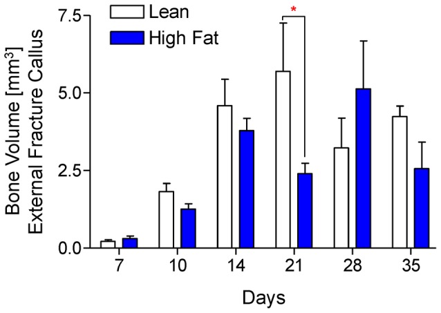
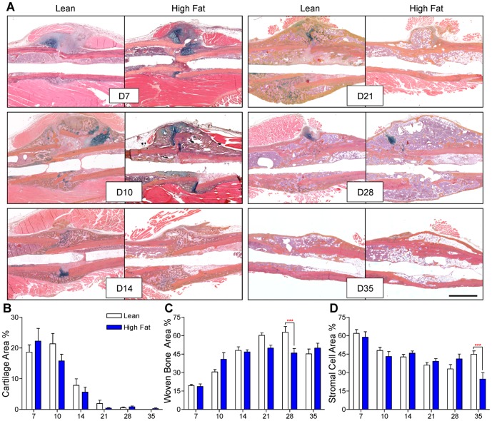
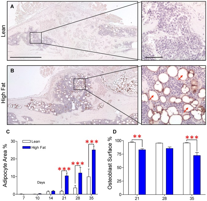
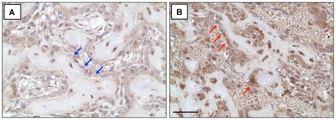
HFD-fed mice displayed increased body weight and impaired glucose tolerance, both characteristic of T2DM. Compared to control mice, HFD-fed mice with tibia fractures showed significantly (p<0.001) decreased woven bone at day 28 by histomorphometry and significantly (p<0.01) decreased callus bone volume at day 21 by μCT. Interestingly, fracture calluses contained markedly increased adiposity in HFD-fed mice at days 21, 28, and 35. HFD-fed mice also showed increased PPARγ immunohistochemical staining at day 14. Finally, calluses from HFD-fed mice at day 35 showed significantly (p<0.01) reduced torsional rigidity compared to controls.
Our murine model of T2DM demonstrated delayed fracture healing and weakened biomechanical properties, and was distinctly characterized by increased callus adiposity. This suggests altered mesenchymal stem cell fate determination with a shift to the adipocyte lineage at the expense of the osteoblast lineage. The up-regulation of PPARγ in fracture calluses of HFD-fed mice is likely involved in the proposed fate switching.
骨骼骨折愈合受损和不愈合是一个重大的公共卫生问题,糖尿病(DM)患者的发病率会加剧。DM在全球范围内普遍存在,影响着约2580万美国成年人,其中超过90%患有肥胖相关的2型糖尿病(T2DM)。虽然已经使用动物模型研究了1型糖尿病(T1DM)中的骨折愈合情况,但尚未对T2DM动物模型中的延迟愈合进行研究。
将5周龄的雄性C57BL/6J小鼠置于对照低脂饮食或实验性高脂饮食(HFD)中12周。在17周龄时诱导胫骨骨干中段开放性骨折,并用脊髓针进行髓内固定。在第7、10、14、21、28和35天处死小鼠,进行微型计算机断层扫描(μCT)、基于组织学的组织形态计量学和分子分析以及生物力学测试。
喂食HFD的小鼠体重增加,葡萄糖耐量受损,这两者都是T2DM的特征。与对照小鼠相比,喂食HFD的胫骨骨折小鼠在第28天时通过组织形态计量学显示编织骨显著减少(p<0.001),在第21天时通过μCT显示骨痂骨体积显著减少(p<0.01)。有趣的是,在第21、28和35天,喂食HFD的小鼠骨折骨痂中的脂肪明显增加。喂食HFD的小鼠在第14天时PPARγ免疫组化染色也增加。最后,与对照组相比,喂食HFD的小鼠在第35天时的骨痂扭转刚度显著降低(p<0.01)。
我们的T2DM小鼠模型显示骨折愈合延迟和生物力学性能减弱,其明显特征是骨痂脂肪增加。这表明间充质干细胞命运决定发生改变,转向脂肪细胞谱系而牺牲成骨细胞谱系。喂食HFD小鼠骨折骨痂中PPARγ的上调可能与所提出的命运转换有关。