Li Yi, Liu Huizhan, Li Jun, Zhang Qian, Gong Shusheng, He David
Department of Otolaryngology-Head and Neck Surgery, Beijing Tongren Hospital, Capital Medical University, Beijing, P.R. China; Department of Biomedical Sciences, Creighton University School of Medicine, Omaha, Nebraska, United States of America.
Department of Biomedical Sciences, Creighton University School of Medicine, Omaha, Nebraska, United States of America.
PLoS One. 2014 Jun 12;9(6):e99840. doi: 10.1371/journal.pone.0099840. eCollection 2014.
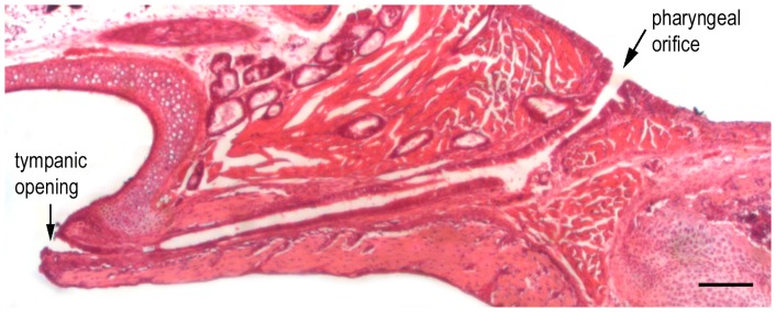
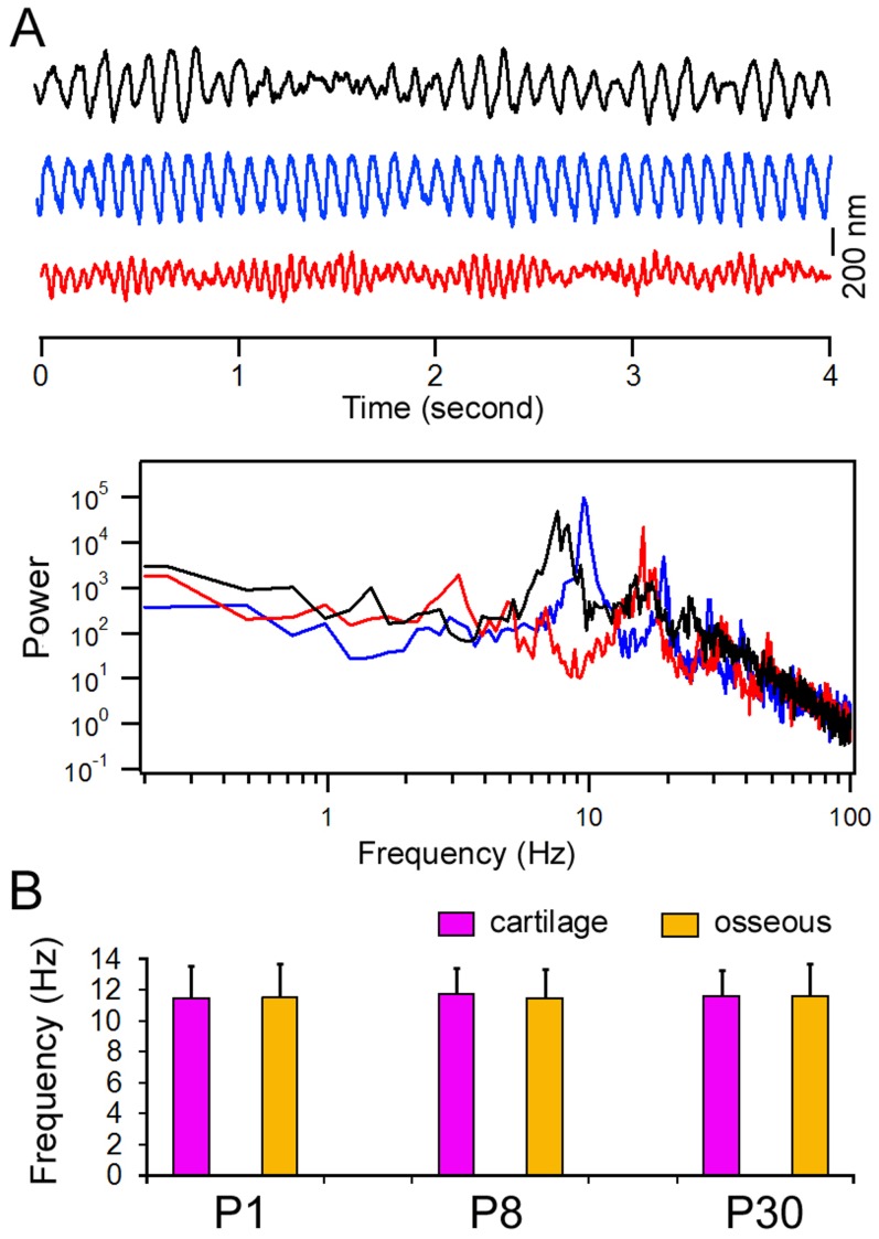
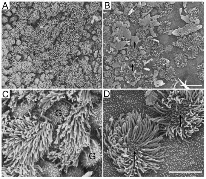
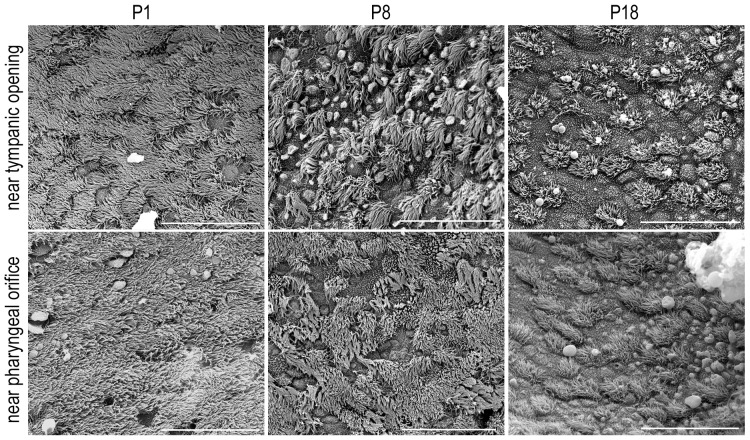
The Eustachian tube is a small canal that connects the tympanic cavity with the nasal part of the pharynx. The epithelial lining of the Eustachian tube contains a ciliated columnar epithelium at the tympanic cavity and a pseudostratified, ciliated columnar epithelium with goblet cells near the pharynx. The tube serves to equalize air pressure across the eardrum and drains mucus away from the middle ear into the nasopharynx. Blockage of the Eustachian tube is the most common cause of all forms of otitis media, which is common in children. In the present study, we examined the epithelial lining of the Eustachian tube in neonatal and adult gerbils, with a focus on the morphological and functional development of ciliated cells in the mucosa. The length of the tube is ∼8.8 mm in adult gerbils. Scanning electron microscopy showed that the mucosal member near the pharyngeal side contains a higher density of ciliated cells and goblet cells than that near the tympanic side. The cilia beat frequency is 11 Hz. During development, the length of the Eustachian tube increased significantly between postnatal day 1 (P1) and P18. Scanning electron microscopy showed that the mucosa contained a high density of ciliated cells with a few goblet cells at P1. The density of ciliated cells decreased while the density of goblet cells increased during development. At P18, the mucosa appeared to be adult-like. Interestingly, the ciliary beat frequency measured from ciliated cells at P1 was not statistically different from that measured from adult animals. Our study suggests that the Eustachian tube undergoes significant anatomical and histological changes between P1 and P18. The tube is morphologically and functionally mature at P18, when the auditory function (sensitivity and frequency selectivity) is mature in this species.
咽鼓管是一条连接鼓室与鼻咽部的小管。咽鼓管的上皮衬里在鼓室处为纤毛柱状上皮,在靠近咽部处为假复层纤毛柱状上皮并含有杯状细胞。该管用于平衡鼓膜两侧的气压,并将中耳的黏液引流至鼻咽部。咽鼓管堵塞是所有类型中耳炎最常见的病因,中耳炎在儿童中很常见。在本研究中,我们检查了新生和成年沙鼠的咽鼓管上皮衬里,重点关注黏膜中纤毛细胞的形态和功能发育。成年沙鼠的咽鼓管长度约为8.8毫米。扫描电子显微镜显示,靠近咽部一侧的黏膜中纤毛细胞和杯状细胞的密度高于靠近鼓室一侧。纤毛摆动频率为11赫兹。在发育过程中,咽鼓管的长度在出生后第1天(P1)到P18之间显著增加。扫描电子显微镜显示,在P1时黏膜中有高密度的纤毛细胞和少量杯状细胞。在发育过程中,纤毛细胞的密度降低而杯状细胞的密度增加。在P18时,黏膜看起来类似成年状态。有趣的是,P1时纤毛细胞测得的纤毛摆动频率与成年动物测得的频率在统计学上无差异。我们的研究表明,咽鼓管在P1到P18之间经历了显著的解剖学和组织学变化。在P18时,当该物种的听觉功能(敏感性和频率选择性)成熟时,咽鼓管在形态和功能上也成熟了。