• 文献检索
  • 文档翻译
  • 深度研究
  • 学术资讯
  • Suppr Zotero 插件Zotero 插件
  • 邀请有礼
  • 套餐&价格
  • 历史记录
应用&插件
Suppr Zotero 插件Zotero 插件浏览器插件Mac 客户端Windows 客户端微信小程序
定价
高级版会员购买积分包购买API积分包
服务
文献检索文档翻译深度研究API 文档MCP 服务
关于我们
关于 Suppr公司介绍联系我们用户协议隐私条款
关注我们

Suppr 超能文献

核心技术专利:CN118964589B侵权必究
粤ICP备2023148730 号-1Suppr @ 2026

文献检索

告别复杂PubMed语法,用中文像聊天一样搜索,搜遍4000万医学文献。AI智能推荐,让科研检索更轻松。

立即免费搜索

文件翻译

保留排版,准确专业,支持PDF/Word/PPT等文件格式,支持 12+语言互译。

免费翻译文档

深度研究

AI帮你快速写综述,25分钟生成高质量综述,智能提取关键信息,辅助科研写作。

立即免费体验

小鼠心脏的心外膜与房室交界区的发育

The Epicardium and the Development of the Atrioventricular Junction in the Murine Heart.

作者信息

Lockhart Marie M, Phelps Aimee L, van den Hoff Maurice J B, Wessels Andy

机构信息

Department of Regenerative Medicine and Cell Biology, Medical University of South Carolina, 173 Ashley Avenue, Charleston, SC 29425, USA;

Academic Medical Center, Heart Failure Research Center, Department of Anatomy, Embryology and Physiology, Meibergdreef 15, 1105AZ, Amsterdam, The Netherlands;

出版信息

J Dev Biol. 2014 Mar 1;2(1):1-17. doi: 10.3390/jdb2010001.

DOI:10.3390/jdb2010001
PMID:24926431
原文链接:https://pmc.ncbi.nlm.nih.gov/articles/PMC4051323/
Abstract

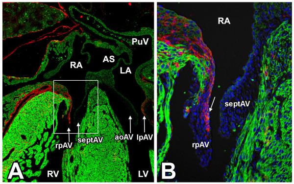
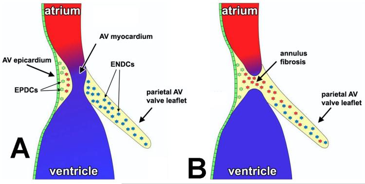
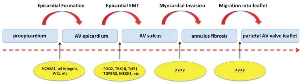
Insight into the role of the epicardium in cardiac development and regeneration has significantly improved over the past ten years. This is mainly due to the increasing availability of new mouse models for the study of the epicardial lineage. Here we focus on the growing understanding of the significance of the epicardium and epicardially-derived cells in the formation of the atrioventricular (AV) junction. First, through the process of epicardial epithelial-to-mesenchymal transformation (epiEMT), the subepicardial AV mesenchyme is formed. Subsequently, the AV-epicardium and epicardially-derived cells (EPDCs) form the annulus fibrosus, a structure important for the electrical separation of atrial and ventricular myocardium. Finally, the AV-EPDCs preferentially migrate into the parietal AV valve leaflets, largely replacing the endocardially-derived cell population. In this review, we provide an overview of what is currently known about the regulation of the events involved in this process.

摘要

在过去十年中,对心外膜在心脏发育和再生中作用的认识有了显著提高。这主要归功于用于研究心外膜谱系的新型小鼠模型越来越容易获得。在这里,我们重点关注对心外膜和心外膜衍生细胞在房室(AV)连接形成中的重要性的日益深入的理解。首先,通过心外膜上皮-间充质转化(epiEMT)过程,形成心外膜下AV间充质。随后,AV-心外膜和心外膜衍生细胞(EPDCs)形成纤维环,这是心房和心室心肌电分离的重要结构。最后,AV-EPDCs优先迁移到房室瓣叶,在很大程度上取代了心内膜衍生的细胞群体。在这篇综述中,我们概述了目前已知的关于该过程中涉及事件的调控情况。

https://cdn.ncbi.nlm.nih.gov/pmc/blobs/345f/4051323/ababaec65689/nihms-591723-f0003.jpg
https://cdn.ncbi.nlm.nih.gov/pmc/blobs/345f/4051323/acac20f8b940/nihms-591723-f0001.jpg
https://cdn.ncbi.nlm.nih.gov/pmc/blobs/345f/4051323/800507bbe6ed/nihms-591723-f0002.jpg
https://cdn.ncbi.nlm.nih.gov/pmc/blobs/345f/4051323/ababaec65689/nihms-591723-f0003.jpg
https://cdn.ncbi.nlm.nih.gov/pmc/blobs/345f/4051323/acac20f8b940/nihms-591723-f0001.jpg
https://cdn.ncbi.nlm.nih.gov/pmc/blobs/345f/4051323/800507bbe6ed/nihms-591723-f0002.jpg
https://cdn.ncbi.nlm.nih.gov/pmc/blobs/345f/4051323/ababaec65689/nihms-591723-f0003.jpg

相似文献

1
The Epicardium and the Development of the Atrioventricular Junction in the Murine Heart.小鼠心脏的心外膜与房室交界区的发育
J Dev Biol. 2014 Mar 1;2(1):1-17. doi: 10.3390/jdb2010001.
2
Alk3 mediated Bmp signaling controls the contribution of epicardially derived cells to the tissues of the atrioventricular junction.Alk3介导的骨形态发生蛋白信号通路控制心外膜来源细胞对房室交界组织的贡献。
Dev Biol. 2014 Dec 1;396(1):8-18. doi: 10.1016/j.ydbio.2014.09.031. Epub 2014 Oct 6.
3
Epicardially derived fibroblasts preferentially contribute to the parietal leaflets of the atrioventricular valves in the murine heart.心外膜衍生的成纤维细胞优先贡献于小鼠心脏房室瓣的心外膜叶。
Dev Biol. 2012 Jun 15;366(2):111-24. doi: 10.1016/j.ydbio.2012.04.020. Epub 2012 Apr 24.
4
The Role of the Epicardium in the Formation of the Cardiac Valves in the Mouse心外膜在小鼠心脏瓣膜形成中的作用
5
Experimental studies on the spatiotemporal expression of WT1 and RALDH2 in the embryonic avian heart: a model for the regulation of myocardial and valvuloseptal development by epicardially derived cells (EPDCs).WT1和RALDH2在鸡胚心脏中的时空表达实验研究:心外膜来源细胞(EPDCs)对心肌和房室间隔发育调控的模型
Dev Biol. 2002 Jul 15;247(2):307-26. doi: 10.1006/dbio.2002.0706.
6
Role of the Epicardium in the Development of the Atrioventricular Valves and Its Relevance to the Pathogenesis of Myxomatous Valve Disease.心外膜在房室瓣发育中的作用及其与黏液瘤样瓣膜病发病机制的相关性。
J Cardiovasc Dev Dis. 2021 May 12;8(5):54. doi: 10.3390/jcdd8050054.
7
Origin, fate, and function of epicardium-derived cells (EPDCs) in normal and abnormal cardiac development.正常和异常心脏发育过程中的心外膜衍生细胞(EPDCs)的起源、命运及功能
ScientificWorldJournal. 2007 Nov 12;7:1777-98. doi: 10.1100/tsw.2007.294.
8
Periostin expression by epicardium-derived cells is involved in the development of the atrioventricular valves and fibrous heart skeleton.心外膜来源细胞的骨膜蛋白表达参与房室瓣和心脏纤维骨架的发育。
Differentiation. 2008 Sep;76(7):809-19. doi: 10.1111/j.1432-0436.2007.00262.x. Epub 2008 Feb 21.
9
Epicardium-derived cells contribute a novel population to the myocardial wall and the atrioventricular cushions.心外膜来源的细胞为心肌壁和房室瓣垫增添了新的细胞群体。
Circ Res. 1998 Jun 1;82(10):1043-52. doi: 10.1161/01.res.82.10.1043.
10
Epicardial deletion of Sox9 leads to myxomatous valve degeneration and identifies Cd109 as a novel gene associated with valve development.心外膜 Sox9 的缺失导致黏液样瓣膜退行性变,并确定 Cd109 为与瓣膜发育相关的新基因。
J Mol Cell Cardiol. 2024 Jan;186:16-30. doi: 10.1016/j.yjmcc.2023.11.002. Epub 2023 Nov 5.

引用本文的文献

1
Foxf1-mediated co-regulation of miR-495 and let-7c modulates epicardial cell migration and myocardial specification.Foxf1介导的miR-495和let-7c的共同调节作用调控心外膜细胞迁移和心肌特化。
Cell Mol Life Sci. 2025 Jun 25;82(1):254. doi: 10.1007/s00018-025-05735-4.
2
Sox9 in the epicardium: Implications for cell invasion, differentiation, and coronary vascular development.心外膜中的Sox9:对细胞侵袭、分化及冠状动脉血管发育的影响
PLoS One. 2025 Jun 23;20(6):e0325852. doi: 10.1371/journal.pone.0325852. eCollection 2025.
3
Rare Variant in Associated With Familial Supraventricular Tachycardia and Wolff-Parkinson-White Syndrome.

本文引用的文献

1
The Wilms' tumor suppressor Wt1 regulates Coronin 1B expression in the epicardium.Wilms 瘤抑制因子 Wt1 调控心外膜中 Coronin 1B 的表达。
Exp Cell Res. 2013 Jun 10;319(10):1365-81. doi: 10.1016/j.yexcr.2013.03.027. Epub 2013 Apr 4.
2
Snai1 is important for avian epicardial cell transformation and motility.Snai1 对于禽类的心外膜细胞的转化和运动非常重要。
Dev Dyn. 2013 Jun;242(6):699-708. doi: 10.1002/dvdy.23967. Epub 2013 Apr 29.
3
The transcription factors Tbx18 and Wt1 control the epicardial epithelial-mesenchymal transition through bi-directional regulation of Slug in murine primary epicardial cells.
与家族性室上性心动过速和预激综合征相关的罕见变异。
Circ Genom Precis Med. 2024 Aug;17(4):e004614. doi: 10.1161/CIRCGEN.124.004614. Epub 2024 Jul 2.
4
Application of neural networks in prenatal diagnosis of atrioventricular septal defect.神经网络在房室间隔缺损产前诊断中的应用。
Transl Pediatr. 2024 Jan 29;13(1):26-37. doi: 10.21037/tp-23-394. Epub 2024 Jan 15.
5
Expression of Stem Cell Niche-Related Biomarkers at the Base of the Human Tricuspid Valve.人三尖瓣根部干细胞龛相关生物标志物的表达。
Stem Cells Dev. 2023 Mar;32(5-6):140-151. doi: 10.1089/scd.2022.0253. Epub 2023 Jan 27.
6
Genomic and physiological analyses of the zebrafish atrioventricular canal reveal molecular building blocks of the secondary pacemaker region.对斑马鱼房室管的基因组和生理学分析揭示了次级起搏区的分子构建模块。
Cell Mol Life Sci. 2021 Oct;78(19-20):6669-6687. doi: 10.1007/s00018-021-03939-y. Epub 2021 Sep 23.
7
How Cardiac Embryology Translates into Clinical Arrhythmias.心脏胚胎学如何转化为临床心律失常。
J Cardiovasc Dev Dis. 2021 Jun 13;8(6):70. doi: 10.3390/jcdd8060070.
8
Role of the Epicardium in the Development of the Atrioventricular Valves and Its Relevance to the Pathogenesis of Myxomatous Valve Disease.心外膜在房室瓣发育中的作用及其与黏液瘤样瓣膜病发病机制的相关性。
J Cardiovasc Dev Dis. 2021 May 12;8(5):54. doi: 10.3390/jcdd8050054.
9
PDGFRα: Expression and Function during Mitral Valve Morphogenesis.血小板衍生生长因子受体α:二尖瓣形态发生过程中的表达与功能
J Cardiovasc Dev Dis. 2021 Mar 13;8(3):28. doi: 10.3390/jcdd8030028.
10
The Mesenchymal Cap of the Atrial Septum and Atrial and Atrioventricular Septation.房间隔的间充质帽与心房及房室间隔形成
J Cardiovasc Dev Dis. 2020 Nov 4;7(4):50. doi: 10.3390/jcdd7040050.
转录因子 Tbx18 和 Wt1 通过双向调控 Slug 控制小鼠原代心外膜细胞的心外膜上皮-间充质转化。
PLoS One. 2013;8(2):e57829. doi: 10.1371/journal.pone.0057829. Epub 2013 Feb 28.
4
Characterization of epicardial-derived cardiac interstitial cells: differentiation and mobilization of heart fibroblast progenitors.心外膜衍生心脏间质细胞的特征:心脏成纤维细胞祖细胞的分化和动员。
PLoS One. 2013;8(1):e53694. doi: 10.1371/journal.pone.0053694. Epub 2013 Jan 18.
5
A molecular and genetic outline of cardiac morphogenesis.心脏形态发生的分子和遗传概述。
Acta Physiol (Oxf). 2013 Apr;207(4):588-615. doi: 10.1111/apha.12061. Epub 2013 Feb 1.
6
The bHLH transcription factor Tcf21 is required for lineage-specific EMT of cardiac fibroblast progenitors.bHLH 转录因子 Tcf21 是心脏成纤维细胞祖细胞谱系特异性 EMT 所必需的。
Development. 2012 Jun;139(12):2139-49. doi: 10.1242/dev.079970. Epub 2012 May 9.
7
Epicardially derived fibroblasts preferentially contribute to the parietal leaflets of the atrioventricular valves in the murine heart.心外膜衍生的成纤维细胞优先贡献于小鼠心脏房室瓣的心外膜叶。
Dev Biol. 2012 Jun 15;366(2):111-24. doi: 10.1016/j.ydbio.2012.04.020. Epub 2012 Apr 24.
8
Nf1 limits epicardial derivative expansion by regulating epithelial to mesenchymal transition and proliferation.NF1 通过调节上皮-间质转化和增殖来限制心外膜衍生扩张。
Development. 2012 Jun;139(11):2040-9. doi: 10.1242/dev.074054. Epub 2012 Apr 25.
9
Distinct compartments of the proepicardial organ give rise to coronary vascular endothelial cells.心外膜原基的不同隔室产生冠状血管内皮细胞。
Dev Cell. 2012 Mar 13;22(3):639-50. doi: 10.1016/j.devcel.2012.01.012.
10
Three-dimensional and molecular analysis of the arterial pole of the developing human heart.人类心脏动脉干的三维和分子分析。
J Anat. 2012 Apr;220(4):336-49. doi: 10.1111/j.1469-7580.2012.01474.x. Epub 2012 Feb 1.