• 文献检索
  • 文档翻译
  • 深度研究
  • 学术资讯
  • Suppr Zotero 插件Zotero 插件
  • 邀请有礼
  • 套餐&价格
  • 历史记录
应用&插件
Suppr Zotero 插件Zotero 插件浏览器插件Mac 客户端Windows 客户端微信小程序
定价
高级版会员购买积分包购买API积分包
服务
文献检索文档翻译深度研究API 文档MCP 服务
关于我们
关于 Suppr公司介绍联系我们用户协议隐私条款
关注我们

Suppr 超能文献

核心技术专利:CN118964589B侵权必究
粤ICP备2023148730 号-1Suppr @ 2026

文献检索

告别复杂PubMed语法,用中文像聊天一样搜索,搜遍4000万医学文献。AI智能推荐,让科研检索更轻松。

立即免费搜索

文件翻译

保留排版,准确专业,支持PDF/Word/PPT等文件格式,支持 12+语言互译。

免费翻译文档

深度研究

AI帮你快速写综述,25分钟生成高质量综述,智能提取关键信息,辅助科研写作。

立即免费体验

高通量、自动化定量分析癫痫性皮质发育不良轻度畸形中的白质神经元。

High-throughput, automated quantification of white matter neurons in mild malformation of cortical development in epilepsy.

机构信息

Department of Clinical and Experimental Epilepsy, UCL Institute of Neurology, Queen Square, WC1N 3BG London, UK.

出版信息

Acta Neuropathol Commun. 2014 Jun 13;2:72. doi: 10.1186/2051-5960-2-72.

DOI:10.1186/2051-5960-2-72
PMID:24927775
原文链接:https://pmc.ncbi.nlm.nih.gov/articles/PMC4229809/
Abstract

INTRODUCTION

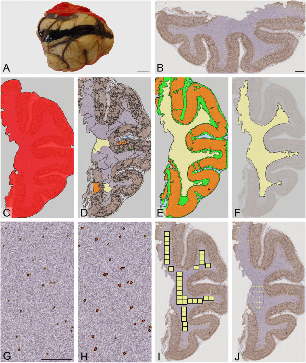
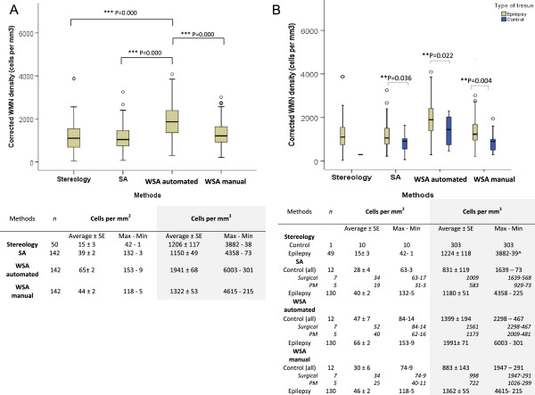
In epilepsy, the diagnosis of mild Malformation of Cortical Development type II (mMCD II) predominantly relies on the histopathological assessment of heterotopic neurons in the white matter. The exact diagnostic criteria for mMCD II are still ill-defined, mainly because findings from previous studies were contradictory due to small sample size, and the use of different stains and quantitative systems. Advance in technology leading to the development of whole slide imaging with high-throughput, automated quantitative analysis (WSA) may overcome these differences, and may provide objective, rapid, and reliable quantitation of white matter neurons in epilepsy. This study quantified the density of NeuN immunopositive neurons in the white matter of up to 142 epilepsy and control cases using WSA. Quantitative data from WSA was compared to two other systems, semi-automated quantitation, and the widely accepted method of stereology, to assess the reliability and quality of results from WSA.

RESULTS

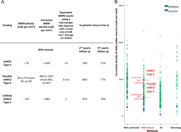
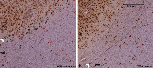
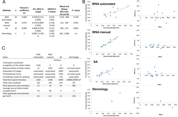
All quantitative systems showed a higher density of white matter neurons in epilepsy cases compared to controls (P = 0.002). We found that, in particular, WSA with user-defined region of interest (manual) was superior in terms of larger sampled size, ease of use, time consumption, and accuracy in region selection and cell recognition compared to other methods. Using results from WSA manual, we proposed a threshold value for the classification of mMCD II, where 78% of patients now classified with mMCD II were seizure-free at the second post-operatively follow up.

CONCLUSION

This study confirms the potential role of WSA in future quantitative diagnostic histology, especially for the histopathological diagnosis of mMCD.

摘要

简介

在癫痫中,轻度皮质发育畸形 II 型(mMCD II)的诊断主要依赖于对脑白质异位神经元的组织病理学评估。mMCD II 的明确诊断标准仍不明确,主要是因为以前的研究由于样本量小,以及使用不同的染色和定量系统,研究结果存在矛盾。技术的进步导致高通量、自动化全切片成像(WSA)的发展,可能克服这些差异,并为癫痫患者脑白质神经元的客观、快速和可靠定量提供可能。本研究使用 WSA 对多达 142 例癫痫和对照病例的脑白质中 NeuN 免疫阳性神经元的密度进行了量化。将 WSA 的定量数据与其他两种系统(半自动定量和广泛接受的体视学法)进行比较,以评估 WSA 结果的可靠性和质量。

结果

所有定量系统均显示癫痫病例的脑白质神经元密度高于对照组(P = 0.002)。我们发现,特别是使用用户定义感兴趣区域的 WSA(手动)在样本量更大、易于使用、耗时和区域选择及细胞识别准确性方面优于其他方法。使用 WSA 手动的结果,我们提出了一个用于 mMCD II 分类的阈值值,其中 78%的被分类为 mMCD II 的患者在第二次术后随访时无癫痫发作。

结论

本研究证实了 WSA 在未来定量诊断组织学中的潜在作用,特别是在 mMCD 的组织病理学诊断中。

https://cdn.ncbi.nlm.nih.gov/pmc/blobs/2421/4229809/5133f4163c0d/40478_2014_139_Fig5_HTML.jpg
https://cdn.ncbi.nlm.nih.gov/pmc/blobs/2421/4229809/f8b203fc7d62/40478_2014_139_Fig1_HTML.jpg
https://cdn.ncbi.nlm.nih.gov/pmc/blobs/2421/4229809/0e8f70e0e64a/40478_2014_139_Fig2_HTML.jpg
https://cdn.ncbi.nlm.nih.gov/pmc/blobs/2421/4229809/bbb967393dbe/40478_2014_139_Fig3_HTML.jpg
https://cdn.ncbi.nlm.nih.gov/pmc/blobs/2421/4229809/99ab5b32cae5/40478_2014_139_Fig4_HTML.jpg
https://cdn.ncbi.nlm.nih.gov/pmc/blobs/2421/4229809/5133f4163c0d/40478_2014_139_Fig5_HTML.jpg

相似文献

1
High-throughput, automated quantification of white matter neurons in mild malformation of cortical development in epilepsy.高通量、自动化定量分析癫痫性皮质发育不良轻度畸形中的白质神经元。
Acta Neuropathol Commun. 2014 Jun 13;2:72. doi: 10.1186/2051-5960-2-72.
2
Changes in vascular density in resected tissue of 97 patients with mild malformation of cortical development, focal cortical dysplasia or TSC-related cortical tubers.97例皮质发育轻度畸形、局灶性皮质发育不良或结节性硬化症相关皮质结节患者切除组织中血管密度的变化。
Int J Dev Neurosci. 2019 Dec;79:96-104. doi: 10.1016/j.ijdevneu.2019.11.003. Epub 2019 Nov 23.
3
Different features of histopathological subtypes of pediatric focal cortical dysplasia.小儿局灶性皮质发育不良组织病理学亚型的不同特征。
Ann Neurol. 2008 Jun;63(6):758-69. doi: 10.1002/ana.21398.
4
[Classification of epileptogenic cerebral malformations: guide to understand the present conditions].[致痫性脑畸形的分类:了解现状指南]
Brain Nerve. 2013 May;65(5):531-40.
5
Dissecting the genetic basis of focal cortical dysplasia: a large cohort study.剖析局灶性皮质发育不良的遗传基础:一项大样本队列研究。
Acta Neuropathol. 2019 Dec;138(6):885-900. doi: 10.1007/s00401-019-02061-5. Epub 2019 Aug 23.
6
Magnetic resonance imaging findings and clinical characteristics in mild malformation of cortical development with oligodendroglial hyperplasia and epilepsy in a predominantly adult cohort.磁共振成像表现及以成胶质细胞增生和癫痫为主要表现的轻度皮质发育不良患者的临床特征,该组患者以成年人为主。
Epilepsia. 2021 Jun;62(6):1429-1441. doi: 10.1111/epi.16907. Epub 2021 Apr 22.
7
Independent contribution of individual white matter pathways to language function in pediatric epilepsy patients.小儿癫痫患者中个体白质通路对语言功能的独立贡献。
Neuroimage Clin. 2014 Sep 30;6:327-32. doi: 10.1016/j.nicl.2014.09.017. eCollection 2014.
8
Neuropathologic measurements in focal cortical dysplasias: validation of the ILAE 2011 classification system and diagnostic implications for MRI.局灶性皮质发育不良的神经病理学测量:对 ILAE 2011 分类系统的验证及其对 MRI 的诊断意义。
Acta Neuropathol. 2012 Feb;123(2):259-72. doi: 10.1007/s00401-011-0920-1. Epub 2011 Nov 27.
9
Age-related MR characteristics in mild malformation of cortical development with oligodendroglial hyperplasia and epilepsy (MOGHE).与少突胶质细胞增生和癫痫相关的轻度皮质发育畸形的年龄相关的 MRI 特征(MOGHE)。
Epilepsy Behav. 2019 Feb;91:68-74. doi: 10.1016/j.yebeh.2018.07.009. Epub 2018 Jul 27.
10
Mild Malformation of Cortical Development with Oligodendroglial Hyperplasia in Frontal Lobe Epilepsy: A New Clinico-Pathological Entity.额叶癫痫中伴有少突胶质细胞增生的轻度皮质发育畸形:一种新的临床病理实体。
Brain Pathol. 2017 Jan;27(1):26-35. doi: 10.1111/bpa.12347. Epub 2016 Feb 22.

引用本文的文献

1
Multiparametric Characterization of Focal Cortical Dysplasia Using 3D MR Fingerprinting.使用三维磁共振指纹技术对焦距性皮质发育异常进行多参数表征
Ann Neurol. 2024 Nov;96(5):944-957. doi: 10.1002/ana.27049. Epub 2024 Aug 3.
2
Combining magnetic resonance fingerprinting with voxel-based morphometric analysis to reduce false positives for focal cortical dysplasia detection.联合磁共振指纹技术与体素形态计量分析减少局灶性皮质发育不良检测的假阳性。
Epilepsia. 2024 Jun;65(6):1631-1643. doi: 10.1111/epi.17951. Epub 2024 Mar 21.
3
Analysis of forensic autopsy cases associated with epilepsy: Comparison between sudden unexpected death in epilepsy (SUDEP) and not-SUDEP groups.

本文引用的文献

1
Comparative expression analysis reveals lineage relationships between human and murine gliomas and a dominance of glial signatures during tumor propagation in vitro.比较表达分析揭示了人类和鼠类神经胶质瘤之间的谱系关系,以及体外肿瘤传播过程中神经胶质特征的主导地位。
Cancer Res. 2013 Sep 15;73(18):5834-44. doi: 10.1158/0008-5472.CAN-13-1299. Epub 2013 Jul 25.
2
Automatic 3D graph cuts for brain cortex segmentation in patients with focal cortical dysplasia.用于局灶性皮质发育异常患者脑皮质分割的自动三维图割技术
Annu Int Conf IEEE Eng Med Biol Soc. 2011;2011:7981-4. doi: 10.1109/IEMBS.2011.6091968.
3
Correlating 3T MRI and histopathology in patients undergoing epilepsy surgery.
与癫痫相关的法医尸检病例分析:癫痫猝死(SUDEP)组与非SUDEP组的比较。
Front Neurol. 2022 Dec 9;13:1077624. doi: 10.3389/fneur.2022.1077624. eCollection 2022.
4
The ILAE consensus classification of focal cortical dysplasia: An update proposed by an ad hoc task force of the ILAE diagnostic methods commission.国际抗癫痫联盟局灶性皮质发育不良分类共识:国际抗癫痫联盟诊断方法委员会特别工作组提出的更新建议。
Epilepsia. 2022 Aug;63(8):1899-1919. doi: 10.1111/epi.17301. Epub 2022 Jun 15.
5
White Matter Interstitial Neurons in the Adult Human Brain: 3% of Cortical Neurons in Quest for Recognition.成人大脑白质间质神经元:占皮质神经元的3%,寻求认可。
Cells. 2021 Jan 19;10(1):190. doi: 10.3390/cells10010190.
6
Can histologically normal epileptogenic zone share common electrophysiological phenotypes with focal cortical dysplasia? SEEG-based study in MRI-negative epileptic patients.组织学正常的致痫区是否与局灶性皮质发育不良具有共同的电生理表型?MRI 阴性癫痫患者的 SEEG 研究。
J Neurol. 2019 Aug;266(8):1907-1918. doi: 10.1007/s00415-019-09339-4. Epub 2019 May 4.
7
Review: The past, present and future challenges in epilepsy-related and sudden deaths and biobanking.综述:癫痫相关和猝死的过去、现在和未来挑战以及生物库。
Neuropathol Appl Neurobiol. 2018 Feb;44(1):32-55. doi: 10.1111/nan.12453.
8
Nestin-expressing cell types in the temporal lobe and hippocampus: Morphology, differentiation, and proliferative capacity.颞叶和海马中的巢蛋白表达细胞类型:形态、分化和增殖能力。
Glia. 2018 Jan;66(1):62-77. doi: 10.1002/glia.23211. Epub 2017 Sep 19.
9
Multinodular and vacuolating neuronal tumors in epilepsy: dysplasia or neoplasia?癫痫性多结节和空泡性神经元肿瘤:发育不良还是肿瘤?
Brain Pathol. 2018 Mar;28(2):155-171. doi: 10.1111/bpa.12555. Epub 2017 Sep 19.
10
Characterising subtypes of hippocampal sclerosis and reorganization: correlation with pre and postoperative memory deficit.描述海马硬化症亚型和重组:与术前和术后记忆缺陷的相关性。
Brain Pathol. 2018 Mar;28(2):143-154. doi: 10.1111/bpa.12514. Epub 2017 Apr 24.
在接受癫痫手术的患者中进行 3T MRI 与组织病理学相关分析。
J Neurosci Methods. 2012 Mar 30;205(1):182-9. doi: 10.1016/j.jneumeth.2011.12.014. Epub 2011 Dec 29.
4
Neuropathologic measurements in focal cortical dysplasias: validation of the ILAE 2011 classification system and diagnostic implications for MRI.局灶性皮质发育不良的神经病理学测量:对 ILAE 2011 分类系统的验证及其对 MRI 的诊断意义。
Acta Neuropathol. 2012 Feb;123(2):259-72. doi: 10.1007/s00401-011-0920-1. Epub 2011 Nov 27.
5
Analysis of protein biomarkers in human clinical tumor samples: critical aspects to success from tissue acquisition to analysis.分析人类临床肿瘤样本中的蛋白质生物标志物:从组织采集到分析成功的关键方面。
Biomark Med. 2011 Apr;5(2):227-48. doi: 10.2217/bmm.11.9.
6
Neuroimaging in identifying focal cortical dysplasia and prognostic factors in pediatric and adolescent epilepsy surgery.神经影像学在儿科和青少年癫痫手术中识别局灶性皮质发育不良和预后因素。
Epilepsia. 2011 Apr;52(4):722-7. doi: 10.1111/j.1528-1167.2010.02950.x. Epub 2011 Jan 28.
7
The clinicopathologic spectrum of focal cortical dysplasias: a consensus classification proposed by an ad hoc Task Force of the ILAE Diagnostic Methods Commission.局灶性皮质发育不良的临床病理谱:国际抗癫痫联盟诊断方法委员会特别工作组提出的共识分类。
Epilepsia. 2011 Jan;52(1):158-74. doi: 10.1111/j.1528-1167.2010.02777.x. Epub 2010 Nov 10.
8
Estimation of nuclear population from microtome sections.从切片估计核数量。
Anat Rec. 1946 Feb;94:239-47. doi: 10.1002/ar.1090940210.
9
Populations of subplate and interstitial neurons in fetal and adult human telencephalon.胎脑和成人端脑基板和间质神经元的群体。
J Anat. 2010 Oct;217(4):381-99. doi: 10.1111/j.1469-7580.2010.01284.x.
10
Developmental history of the subplate zone, subplate neurons and interstitial white matter neurons: relevance for schizophrenia.板下层区域、板下神经元和间质白质神经元的发育史:与精神分裂症的相关性
Int J Dev Neurosci. 2011 May;29(3):193-205. doi: 10.1016/j.ijdevneu.2010.09.005. Epub 2010 Sep 29.