Cole Eye Institute, Cleveland Clinic, Cleveland, Ohio, USA.
Br J Ophthalmol. 2014 Jul;98 Suppl 2(Suppl 2):ii24-9. doi: 10.1136/bjophthalmol-2014-305305.
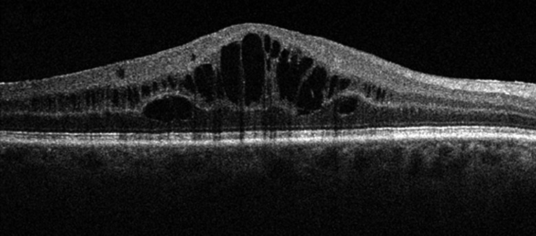
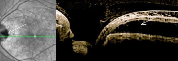
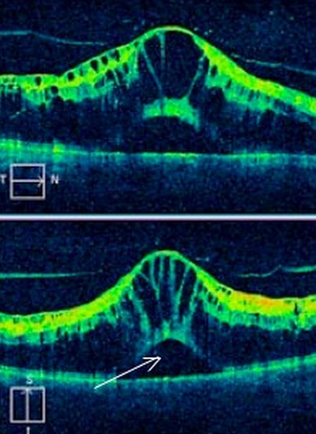
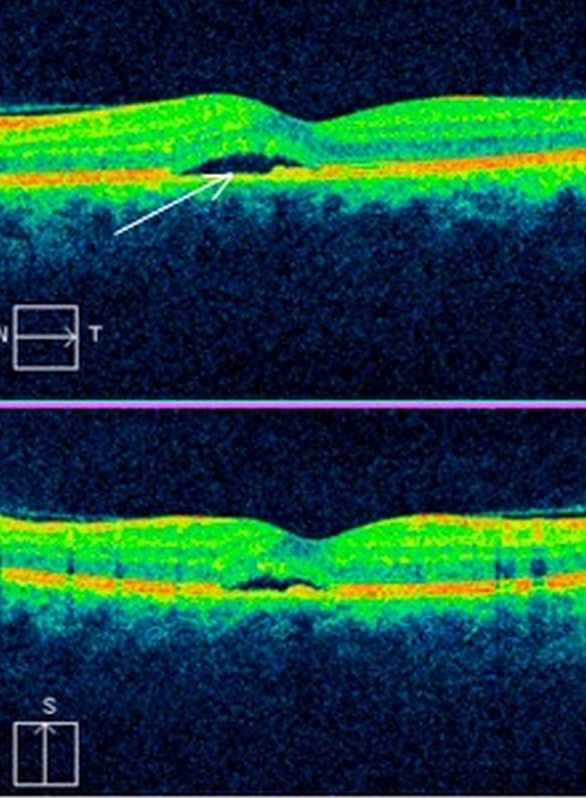
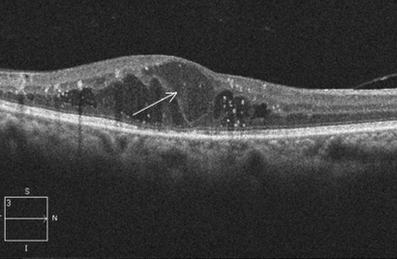
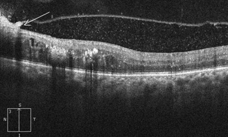
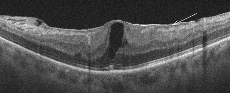
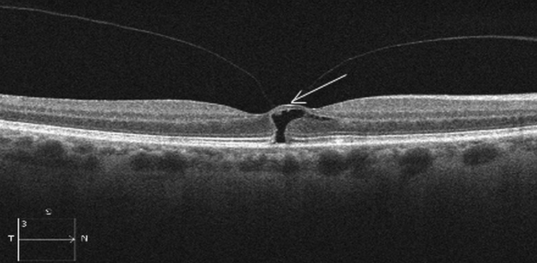
Macular oedema (ME) occurs in a wide variety of pathological conditions and accounts for different degrees of vision loss. Early detection of ME is therefore critical for diagnosis and therapeutic management. Optical coherence tomography (OCT) is a non-contact, diagnostic method that uses infrared light, which allows the analysis of the retinal structure by means of high-resolution tomographic cross sections. The identification, localisation, quantification and long-term follow-up of fluid collections are the most important capabilities of OCT. Since the introduction of OCT in clinical practice, it has become an invaluable diagnostic tool and different patterns of ME have been reported. The purpose of this manuscript is to review OCT profiles of ME according to the aetiology and describe what has been reported regarding intraretinal features in vivo.
黄斑水肿(ME)可发生于多种病理情况,导致不同程度的视力丧失。因此,早期发现 ME 对于诊断和治疗管理至关重要。光学相干断层扫描(OCT)是一种非接触性诊断方法,使用近红外光,可以通过高分辨率断层扫描进行视网膜结构分析。OCT 的最重要功能是识别、定位、量化和长期随访液体积聚。自从 OCT 在临床实践中引入以来,它已成为一种非常有价值的诊断工具,并且已经报道了不同类型的 ME。本文的目的是根据病因回顾 ME 的 OCT 特征,并描述体内已报道的关于视网膜内特征的内容。