• 文献检索
  • 文档翻译
  • 深度研究
  • 学术资讯
  • Suppr Zotero 插件Zotero 插件
  • 邀请有礼
  • 套餐&价格
  • 历史记录
应用&插件
Suppr Zotero 插件Zotero 插件浏览器插件Mac 客户端Windows 客户端微信小程序
定价
高级版会员购买积分包购买API积分包
服务
文献检索文档翻译深度研究API 文档MCP 服务
关于我们
关于 Suppr公司介绍联系我们用户协议隐私条款
关注我们

Suppr 超能文献

核心技术专利:CN118964589B侵权必究
粤ICP备2023148730 号-1Suppr @ 2026

文献检索

告别复杂PubMed语法,用中文像聊天一样搜索,搜遍4000万医学文献。AI智能推荐,让科研检索更轻松。

立即免费搜索

文件翻译

保留排版,准确专业,支持PDF/Word/PPT等文件格式,支持 12+语言互译。

免费翻译文档

深度研究

AI帮你快速写综述,25分钟生成高质量综述,智能提取关键信息,辅助科研写作。

立即免费体验

对比增强能量多普勒内镜超声与超声弹性成像技术应变比值对胰腺局灶性实性病变的诊断价值。

Yield of Contrast-Enhanced Power Doppler Endoscopic Ultrasonography and Strain Ratio Obtained by EUS-Elastography in the Diagnosis of Focal Pancreatic Solid Lesions.

机构信息

Unité d'Exploration Médico-Chirurgicale Oncologique, Institut Paoli-Calmettes, France ; Gastroenterology Department, University of the State of Rio de Janeiro and Federal University of Rio de Janeiro, Brazil.

Biopathology Unit, Institut Paoli-Calmettes, France.

出版信息

Endosc Ultrasound. 2012 Oct;1(3):143-9. doi: 10.7178/eus.03.005.

DOI:10.7178/eus.03.005
PMID:24949352
原文链接:https://pmc.ncbi.nlm.nih.gov/articles/PMC4062225/
Abstract

OBJECTIVE

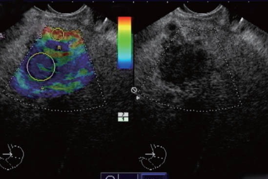
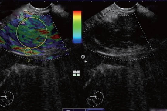
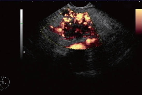
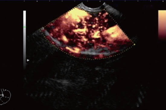
Although endoscopic ultrasonography-guided fine needle aspiration (EUS-FNA) is the gold standard for diagnosing pancreatic lesions, its negative predictive value is suboptimal. Our aim was to evaluate the yield of contrast-enhanced EUS (CED-EUS) and of strain ratio EUS-elastography (SR-E-EUS) for differentiating pancreatic solid lesions.

METHODS

Forty-seven patients (27 men, 20 women, 70 ± 11 years) were consecutively involved in this single-center, prospective study. They were submitted to EUS, SR-E-EUS, CED-EUS with Sonovue(®), and EUS-FNA. The final diagnosis was based on the histological assessment of EUS-FNA and/or surgical specimens when available, and on follow-up of at least 6 months.

RESULTS

From the 47 focal pancreatic lesions included, 13 (28%) were benign and 34 (72%) malignant. Patients with malignancy were older (70 ± 11 vs. 61 ± 8, P = 0.003), and had larger lesions (34 ± 12 mm vs. 22 ± 11 mm, P = 0.03). Malignant lesions had higher SR-E-EUS (31 ± 32 vs. 8 ± 9, P = 0.001) and more hypovascular pattern (93% vs. 33%, P < 0.001). Logistic regression determined that only hypovascularity (OR = 2.6, 95%CI: 1.5-130, P = 0.02) was independently predictive of malignancy. ROC analysis for SR-E-EUS yielded an optimal cutoff of 8 (AUC 0.91, 95%CI: 0.74-0.98) for the best power distinction for malignancy. There was no significant difference concerning sensitivity (79%, 90%, 93%) and specificity rates (85%, 75%, 67%) of EUS-FNA, SR-E-EUS, and CED-EUS, respectively. By analysis of the inconclusive EUS-FNA subset (9 patients, 19%), SR-E-EUS > 8 and hypovascularity showed sensitivity of 80% and 100%, and specificity of 67% and 67%, respectively.

CONCLUSION

The clinical utility of CED-EUS and SR-E-EUS remains questionable. The accuracies of CED-EUS and SR-E-EUS are similar to EUS-FNA. Hypovascularity was independently predictive of malignancy. Patients with inconclusive EUS-FNA could benefit from CED-EUS due to the high sensitivity of hypovascularity for diagnosing malignancy.

摘要

目的

尽管内镜超声引导下细针抽吸术(EUS-FNA)是诊断胰腺病变的金标准,但它的阴性预测值并不理想。我们的目的是评估对比增强超声(CED-EUS)和应变比超声弹性成像(SR-E-EUS)在鉴别胰腺实性病变方面的应用价值。

方法

47 例患者(男 27 例,女 20 例,70±11 岁)连续参与了这项单中心前瞻性研究。他们接受了 EUS、SR-E-EUS、CEDEUS 联合 SonoVue(®)和 EUS-FNA。最终诊断基于 EUS-FNA 的组织学评估和/或手术标本,如有,并进行至少 6 个月的随访。

结果

47 个局灶性胰腺病变中,13 个(28%)为良性,34 个(72%)为恶性。恶性肿瘤患者年龄更大(70±11 岁 vs. 61±8 岁,P=0.003),病变更大(34±12 毫米 vs. 22±11 毫米,P=0.03)。恶性病变的 SR-E-EUS 值更高(31±32 与 8±9,P=0.001),且更倾向于低血流灌注模式(93%与 33%,P<0.001)。Logistic 回归分析确定,只有低血流灌注(OR=2.6,95%CI:1.5-130,P=0.02)是恶性肿瘤的独立预测因素。SR-E-EUS 的 ROC 分析得到了一个最佳的截断值为 8(AUC 0.91,95%CI:0.74-0.98),以区分良恶性病变。EUS-FNA、SR-E-EUS 和 CED-EUS 的诊断恶性肿瘤的敏感性(分别为 79%、90%和 93%)和特异性(分别为 85%、75%和 67%)差异无统计学意义。通过对 EUS-FNA 不确定的亚组(9 例,19%)进行分析,SR-E-EUS>8 和低血流灌注的敏感性分别为 80%和 100%,特异性分别为 67%和 67%。

结论

CED-EUS 和 SR-E-EUS 的临床应用价值仍存在疑问。CED-EUS 和 SR-E-EUS 的准确性与 EUS-FNA 相似。低血流灌注是恶性肿瘤的独立预测因素。EUS-FNA 不确定的患者可以从 CED-EUS 中受益,因为低血流灌注对诊断恶性肿瘤的敏感性很高。

https://cdn.ncbi.nlm.nih.gov/pmc/blobs/8eab/4062225/210891a14dd9/EUS-1-143-g006.jpg
https://cdn.ncbi.nlm.nih.gov/pmc/blobs/8eab/4062225/7f53b731efa6/EUS-1-143-g001.jpg
https://cdn.ncbi.nlm.nih.gov/pmc/blobs/8eab/4062225/c1bd50db3dfc/EUS-1-143-g002.jpg
https://cdn.ncbi.nlm.nih.gov/pmc/blobs/8eab/4062225/06a637464fc8/EUS-1-143-g003.jpg
https://cdn.ncbi.nlm.nih.gov/pmc/blobs/8eab/4062225/9be61e8b1e1d/EUS-1-143-g004.jpg
https://cdn.ncbi.nlm.nih.gov/pmc/blobs/8eab/4062225/210891a14dd9/EUS-1-143-g006.jpg

相似文献

1
Yield of Contrast-Enhanced Power Doppler Endoscopic Ultrasonography and Strain Ratio Obtained by EUS-Elastography in the Diagnosis of Focal Pancreatic Solid Lesions.对比增强能量多普勒内镜超声与超声弹性成像技术应变比值对胰腺局灶性实性病变的诊断价值。
Endosc Ultrasound. 2012 Oct;1(3):143-9. doi: 10.7178/eus.03.005.
2
Combination of EUS-FNA and elastography (strain ratio) to exclude malignant solid pancreatic lesions: A prospective single-blinded study.超声内镜引导下细针穿刺活检(EUS-FNA)与弹性成像(应变率)联合应用以排除胰腺实性恶性病变:一项前瞻性单盲研究。
J Gastroenterol Hepatol. 2015 Nov;30(11):1683-9. doi: 10.1111/jgh.13067.
3
[Value of endoscopic ultrasound-guided fine needle aspiration in pretest prediction and diagnosis of pancreatic ductal adenocarcinoma].[内镜超声引导下细针穿刺在胰腺导管腺癌术前预测及诊断中的价值]
Nan Fang Yi Ke Da Xue Xue Bao. 2018 Sep 30;38(10):1171-1178. doi: 10.3969/j.issn.1673-4254.2018.10.04.
4
Characterization of solid focal pancreatic lesions using endoscopic ultrasonography with real-time elastography.使用内镜超声结合实时弹性成像对胰腺实性局灶性病变进行特征分析。
Scand J Gastroenterol. 2014 Jun;49(6):742-51. doi: 10.3109/00365521.2014.905627. Epub 2014 Apr 8.
5
Prospective cohort study comparing transient EUS guided elastography to EUS-FNA for the diagnosis of solid pancreatic mass lesions.比较经内镜超声引导下瞬时弹性成像与内镜超声引导下细针穿刺活检诊断胰腺实性肿块病变的前瞻性队列研究。
Pancreatology. 2016 Jan-Feb;16(1):110-4. doi: 10.1016/j.pan.2015.10.003. Epub 2015 Nov 2.
6
Conventional versus contrast-enhanced harmonic endoscopic ultrasonography-guided fine-needle aspiration for diagnosis of solid pancreatic lesions: A prospective randomized trial.传统超声内镜引导下细针穿刺与对比增强谐波超声内镜引导下细针穿刺诊断胰腺实性病变的前瞻性随机试验
Pancreatology. 2015 Sep-Oct;15(5):538-541. doi: 10.1016/j.pan.2015.06.005. Epub 2015 Jun 24.
7
Increasing Number of Passes Beyond 4 Does Not Increase Sensitivity of Detection of Pancreatic Malignancy by Endoscopic Ultrasound-Guided Fine-Needle Aspiration.内镜超声引导下细针抽吸术超过 4 次的穿刺次数增加并不会提高胰腺恶性肿瘤的检出率。
Clin Gastroenterol Hepatol. 2017 Jul;15(7):1071-1078.e2. doi: 10.1016/j.cgh.2016.12.018. Epub 2016 Dec 23.
8
Yield of endoscopic ultrasound-guided fine-needle aspiration of bile duct lesions.内镜超声引导下胆管病变细针穿刺活检的取材率
Endoscopy. 2004 Aug;36(8):715-9. doi: 10.1055/s-2004-825657.
9
Relationship of pancreatic mass size and diagnostic yield of endoscopic ultrasound-guided fine needle aspiration.胰腺肿块大小与超声内镜引导下细针抽吸诊断率的关系。
Dig Dis Sci. 2011 Nov;56(11):3370-5. doi: 10.1007/s10620-011-1782-z. Epub 2011 Jun 19.
10
The Role of Real Time Endoscopic Ultrasound Guided Elastography for Targeting EUS-FNA of Suspicious Pancreatic Masses: A Review of the Literature and A Single Center Experience.实时内镜超声引导弹性成像在可疑胰腺肿块EUS-FNA靶向中的作用:文献综述及单中心经验
JOP. 2016 Sep;17(5):516-524.

引用本文的文献

1
Endoscopic Ultrasound (EUS) Guided Elastography.内镜超声(EUS)引导下弹性成像
Diagnostics (Basel). 2023 May 10;13(10):1686. doi: 10.3390/diagnostics13101686.
2
Advances in Pediatric Diagnostic Endoscopy: A State-of-the-Art Review.儿科诊断性内镜检查的进展:一项最新综述。
JPGN Rep. 2022 Jul 29;3(3):e224. doi: 10.1097/PG9.0000000000000224. eCollection 2022 Aug.
3
Contrast Enhanced EUS for Predicting Solid Pancreatic Neuroendocrine Tumor Grade and Aggressiveness.对比增强超声内镜用于预测实性胰腺神经内分泌肿瘤的分级和侵袭性

本文引用的文献

1
Pancreatico-biliary endoscopic ultrasound: a systematic review of the levels of evidence, performance and outcomes.胰腺胆道内镜超声:证据水平、性能和结果的系统评价。
World J Gastroenterol. 2012 Aug 28;18(32):4243-56. doi: 10.3748/wjg.v18.i32.4243.
2
Diagnostic accuracy of quantitative EUS elastography for discriminating malignant from benign solid pancreatic masses: a prospective, single-center study.定量超声弹性成像诊断胰腺实性肿块良恶性的准确性:一项前瞻性单中心研究。
Gastrointest Endosc. 2012 Nov;76(5):953-61. doi: 10.1016/j.gie.2012.05.034. Epub 2012 Jul 31.
3
Efficacy of an artificial neural network-based approach to endoscopic ultrasound elastography in diagnosis of focal pancreatic masses.
Diagnostics (Basel). 2023 Jan 9;13(2):239. doi: 10.3390/diagnostics13020239.
4
The efficiency of contrast-enhanced endoscopic ultrasound (EUS) combined with EUS elastography for pancreatic cancer diagnosis: a systematic review and meta-analysis.对比增强内镜超声(EUS)联合EUS弹性成像在胰腺癌诊断中的效能:一项系统评价和Meta分析
Ultrasonography. 2023 Jan;42(1):20-30. doi: 10.14366/usg.22103. Epub 2022 Oct 26.
5
Current role of endoscopic ultrasound in the diagnosis and management of pancreatic cancer.内镜超声在胰腺癌诊断和治疗中的当前作用。
World J Gastrointest Endosc. 2022 Jan 16;14(1):35-48. doi: 10.4253/wjge.v14.i1.35.
6
Recent Advances in Endosonography-Elastography: Literature Review.内镜超声弹性成像的最新进展:文献综述
J Clin Med. 2021 Aug 23;10(16):3739. doi: 10.3390/jcm10163739.
7
Role of Endoscopic Ultrasonography and Endoscopic Retrograde Cholangiopancreatography in the Diagnosis of Pancreatic Cancer.内镜超声检查和内镜逆行胰胆管造影术在胰腺癌诊断中的作用
Diagnostics (Basel). 2021 Feb 4;11(2):238. doi: 10.3390/diagnostics11020238.
8
The role of endobronchial ultrasonography elastography for predicting malignancy.支气管内超声弹性成像在预测恶性肿瘤方面的作用。
Turk Gogus Kalp Damar Cerrahisi Derg. 2020 Jan 23;28(1):158-165. doi: 10.5606/tgkdc.dergisi.2020.18508. eCollection 2020 Jan.
9
Impact of endoscopic ultrasonography on diagnosis of pancreatic cancer.内镜超声检查对胰腺癌诊断的影响。
J Gastroenterol. 2019 Jan;54(1):19-32. doi: 10.1007/s00535-018-1519-2. Epub 2018 Nov 7.
10
Endoscopic ultrasound (EUS) elastography and strain ratio, could it help in differentiating malignant from benign pancreatic lesions?内镜超声(EUS)弹性成像和应变率,能否有助于鉴别胰腺良恶性病变?
Medicine (Baltimore). 2018 Sep;97(36):e11689. doi: 10.1097/MD.0000000000011689.
基于人工神经网络的内镜超声弹性成像在诊断局灶性胰腺肿块中的效能。
Clin Gastroenterol Hepatol. 2012 Jan;10(1):84-90.e1. doi: 10.1016/j.cgh.2011.09.014. Epub 2011 Oct 1.
4
Combined contrast-enhanced power Doppler and real-time sonoelastography performed during EUS, used in the differential diagnosis of focal pancreatic masses (with videos).超声内镜检查时联合使用增强能量多普勒和实时超声弹性成像进行诊断,用于鉴别诊断局灶性胰腺肿块(附有视频)。
Gastrointest Endosc. 2010 Oct;72(4):739-47. doi: 10.1016/j.gie.2010.02.056. Epub 2010 Aug 2.
5
Quantitative endoscopic ultrasound elastography: an accurate method for the differentiation of solid pancreatic masses.定量内镜超声弹性成像:一种准确区分胰腺实性肿块的方法。
Gastroenterology. 2010 Oct;139(4):1172-80. doi: 10.1053/j.gastro.2010.06.059. Epub 2010 Jun 27.
6
Contrast-enhanced harmonic endoscopic ultrasound in solid lesions of the pancreas: results of a pilot study.超声内镜引导下胰腺实性病灶造影增强谐波成像:一项初步研究结果。
Endoscopy. 2010 Jul;42(7):564-70. doi: 10.1055/s-0030-1255537. Epub 2010 Jun 30.
7
Pancreatic endocrine tumors: a large single-center experience.胰腺内分泌肿瘤:一项大型单中心经验。
Pancreas. 2009 Nov;38(8):936-40. doi: 10.1097/MPA.0b013e3181b365db.
8
Indications and limitations of endoscopic ultrasound elastography for evaluation of focal pancreatic lesions.内镜超声弹性成像在评估胰腺局灶性病变中的适应证与局限性
Endoscopy. 2008 Nov;40(11):910-7. doi: 10.1055/s-2008-1077726. Epub 2008 Nov 13.
9
Improved differentiation of pancreatic tumors using contrast-enhanced endoscopic ultrasound.使用对比增强内镜超声改善胰腺肿瘤的鉴别诊断
Clin Gastroenterol Hepatol. 2008 May;6(5):590-597.e1. doi: 10.1016/j.cgh.2008.02.030.
10
A novel perfusion imaging technique of the pancreas: contrast-enhanced harmonic EUS (with video).一种新型的胰腺灌注成像技术:对比增强谐波内镜超声(附视频)。
Gastrointest Endosc. 2008 Jan;67(1):141-50. doi: 10.1016/j.gie.2007.07.045.