Bai Ying-Ying, Gao Xihui, Wang Yuan-Cheng, Peng Xin-Gui, Chang Di, Zheng Shuyan, Li Cong, Ju Shenghong
1. Jiangsu Key Laboratory of Molecular and Functional Imaging, Department of Radiology, Zhongda Hospital, Medical School, Southeast University, Nanjing, 210009, China;
2. Key Laboratory of Smart Drug Delivery, Ministry of Education & PLA, School of Pharmacy, Fudan University, Shanghai, 201203, China.
Theranostics. 2014 May 25;4(8):787-97. doi: 10.7150/thno.9525. eCollection 2014.
The efficacy of pro-angiogenic therapy is difficult to evaluate with current diagnostic modalities. The objectives were to develop a non-invasive imaging strategy to define the temporal characteristics of angiogenesis and to evaluate the response to pro-angiogenic therapy in diabetic stroke mouse models.
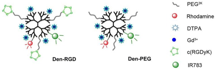
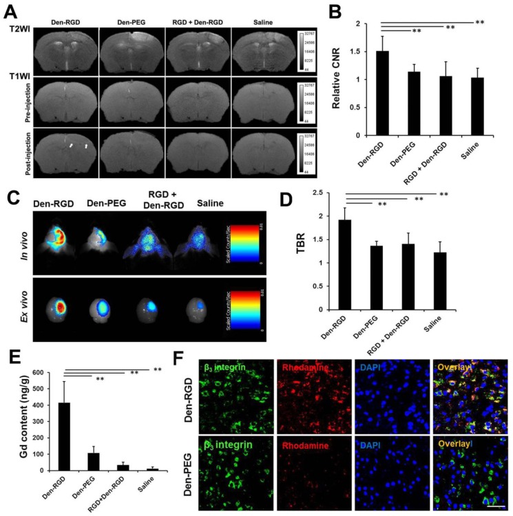
A home-made ανβ3 integrin-targeted multi-modal nanoprobe was intravenously injected into mouse models at set time points after photothrombotic stroke. Magnetic resonance imaging (MRI) and near-infrared fluorescence (NIRF) imaging were carried out at 24 h post-injection. Bone marrow-derived endothelial progenitor cells (EPCs) were infused into the mouse models of ischemic stroke to stimulate angiogenesis.
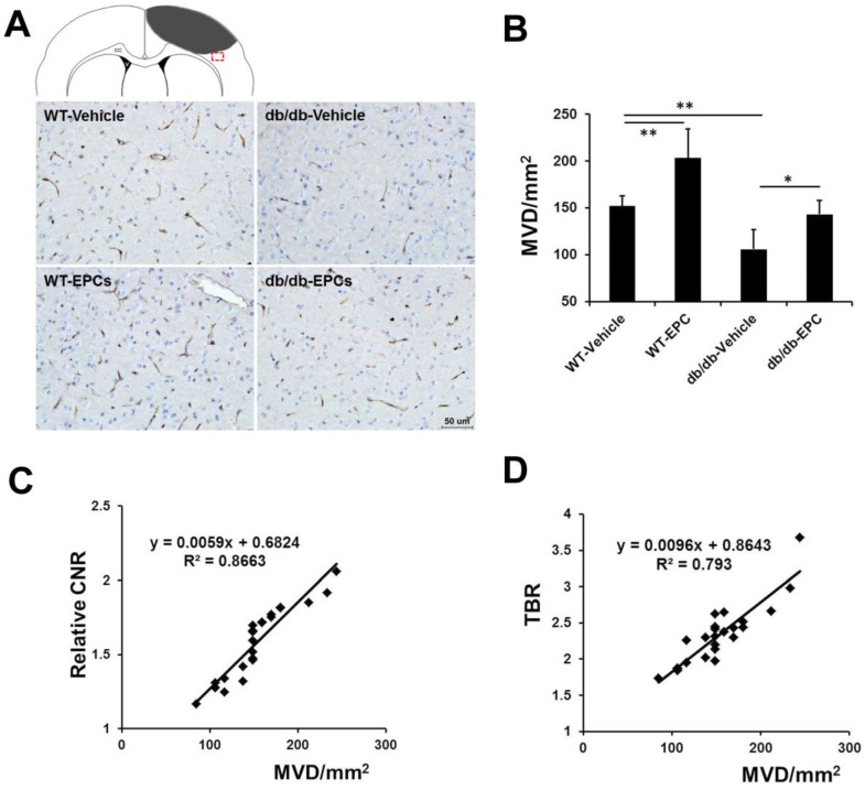
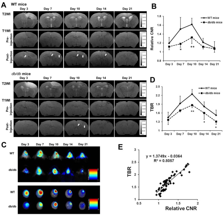
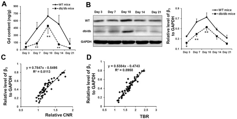
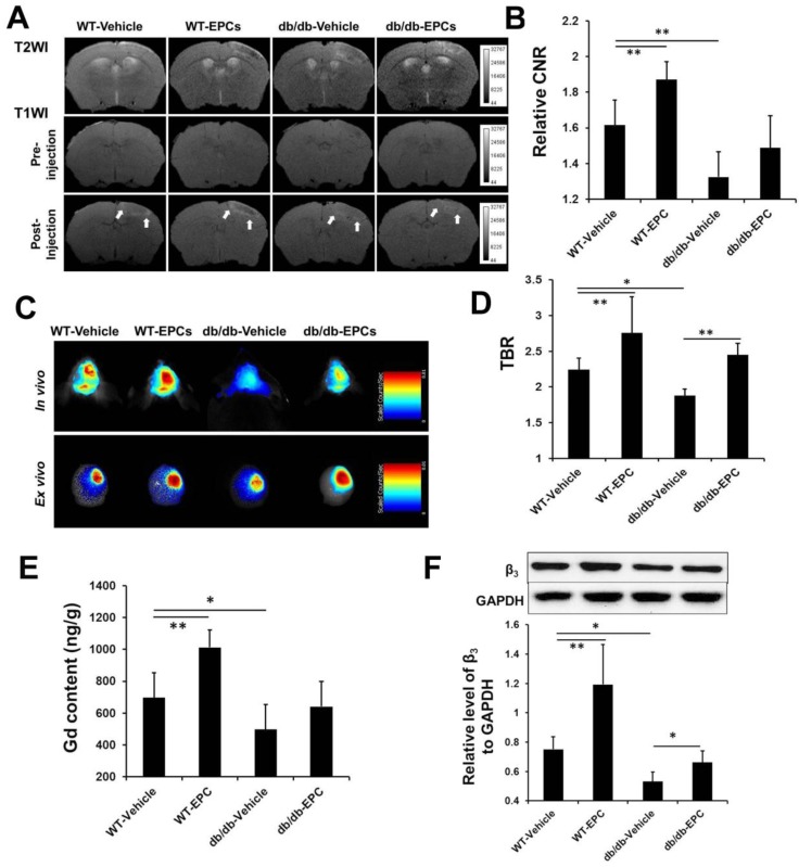
The peak signal intensity in the ischemic-angiogenic area of diabetic and wild-type mouse models was achieved on day 10, with significantly lower signal enhancement observed in the diabetic models. Although the signal intensity was significantly higher after EPC treatment in both models, the enhancement was less pronounced in the diabetic animals compared with the wild-type controls. Histological analysis revealed that the microvessel density and expression of β3 integrin were correlated with the signal intensity assessed with MRI and NIRF imaging.
The non-invasive imaging method could be used for early and accurate evaluation of the response to pro-angiogenic therapy in diabetic stroke models.
目前的诊断方式难以评估促血管生成疗法的疗效。本研究旨在开发一种非侵入性成像策略,以确定血管生成的时间特征,并评估糖尿病性脑卒中小鼠模型对促血管生成疗法的反应。
在光血栓性脑卒中后的设定时间点,将自制的 ανβ3 整合素靶向多模态纳米探针静脉注射到小鼠模型中。注射后 24 小时进行磁共振成像(MRI)和近红外荧光(NIRF)成像。将骨髓源性内皮祖细胞(EPCs)注入缺血性脑卒中小鼠模型以刺激血管生成。
糖尿病和野生型小鼠模型缺血性血管生成区域的信号强度峰值在第 10 天出现,糖尿病模型中的信号增强明显较低。尽管两种模型在 EPC 治疗后信号强度均显著升高,但与野生型对照相比,糖尿病动物的增强作用不那么明显。组织学分析显示,微血管密度和 β3 整合素的表达与通过 MRI 和 NIRF 成像评估的信号强度相关。
这种非侵入性成像方法可用于早期、准确评估糖尿病性脑卒中模型对促血管生成疗法的反应。