Affiliations of authors: University of Texas Graduate School of Biomedical Sciences at Houston, Houston, TX (GV-T, LPh, FZ, P-CC, J-HS, HHC, CG, CC, FJE, M-HL, S-CJY); Cancer Biology Graduate Program (GV-T, P-CC, J-HS, HHC, CC, M-HL), Genes and Development Graduate Program (CG, M-HL); Department of Molecular and Cellular Oncology (EF-M, GV-T, LPh, FZ, P-CC, J-HS, HHC, RZ, JC, CG, CC, FJE, M-HL), Department of Breast Medical Oncology (FJE, GNH, LPu); Department of Biostatistics (JE), Department of Bioinformatics and Computational Biology (YQ); Department of Pathology (YZ, YW, WFS); Department of Emergency Medicine (J-SC, S-CJY), and Department of Endocrine Neoplasia and Hormonal Disorders (S-CJY), The University of Texas MD Anderson Cancer Center, Houston, TX; Center for Cancer & Stem Cell Biology, Institute of Biosciences and Technology, Texas A&M Health Science Center, Houston, TX (YL, WLM); Present address: Breast Cancer Program, Yale Cancer Center, New Haven, CT (LPu).
J Natl Cancer Inst. 2014 Jun 23;106(7). doi: 10.1093/jnci/dju158. Print 2014 Jul.
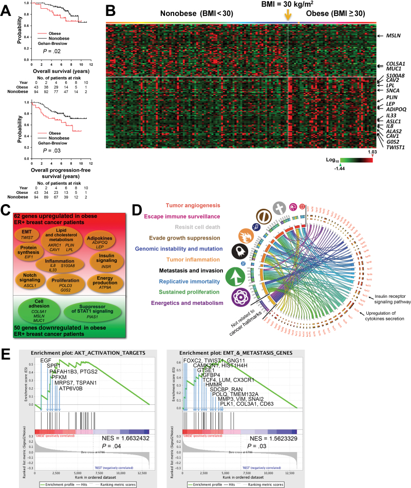
Obesity increases the risk of cancer death among postmenopausal women with estrogen receptor-positive (ER+) breast cancer, but the direct evidence for the mechanisms is lacking. The purpose of this study is to demonstrate direct evidence for the mechanisms mediating this epidemiologic phenomenon.
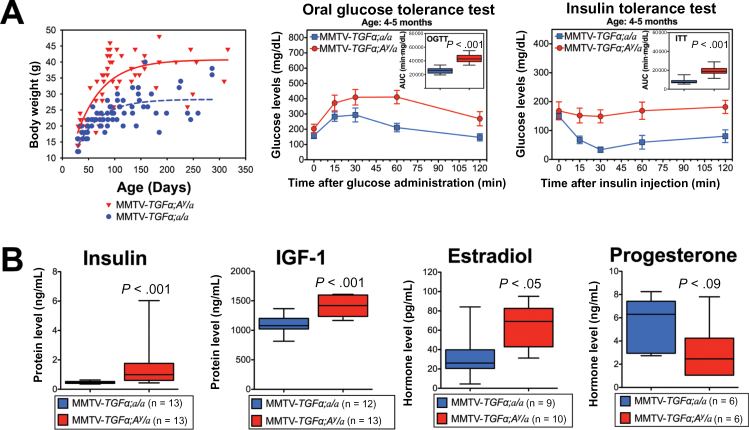
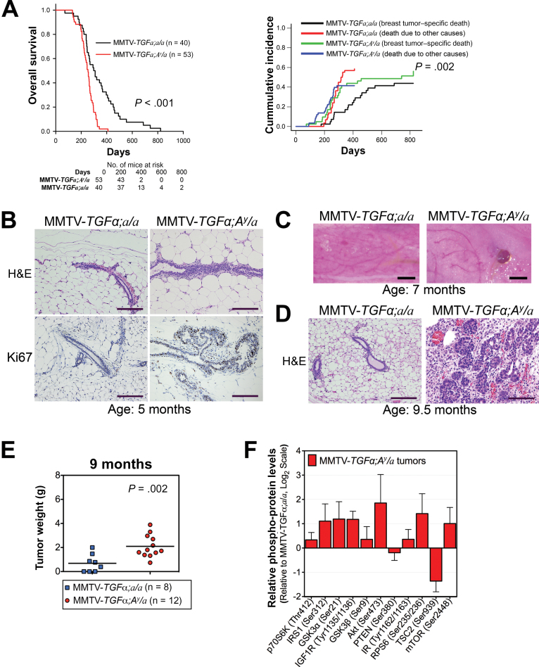
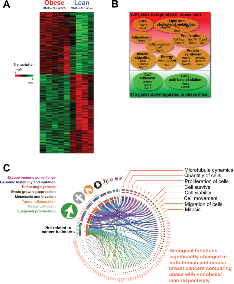
We analyzed transcriptomic profiles of pretreatment biopsies from a prospective cohort of 137 ER+ breast cancer patients. We generated transgenic (MMTV-TGFα;A (y) /a) and orthotopic/syngeneic (A (y) /a) obese mouse models to investigate the effect of obesity on tumorigenesis and tumor progression and to determine biological mechanisms using whole-genome transcriptome microarrays and protein analyses. We used a coculture system to examine the impact of adipocytes/adipokines on breast cancer cell proliferation. All statistical tests were two-sided.
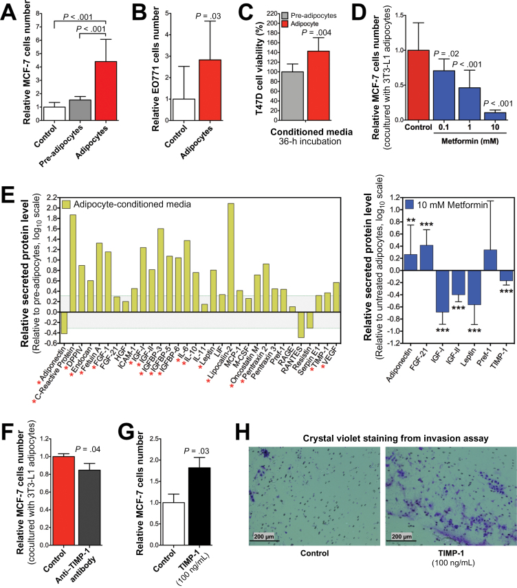
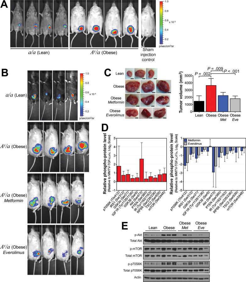
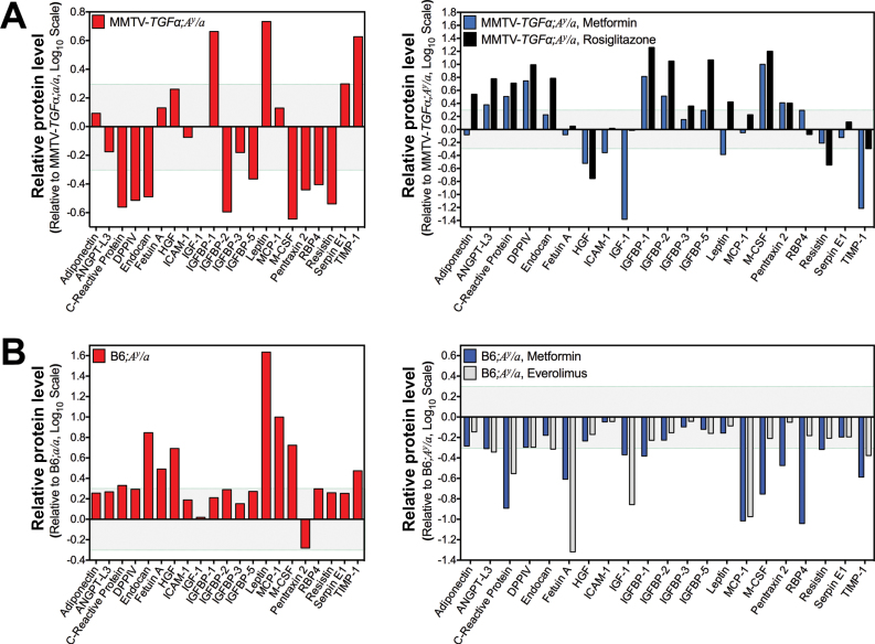
Functional transcriptomic analysis of patients revealed the association of obesity with 59 biological functional changes (P < .05) linked to cancer hallmarks. Gene enrichment analysis revealed enrichment of AKT-target genes (P = .04) and epithelial-mesenchymal transition genes (P = .03) in patients. Our obese mouse models demonstrated activation of the AKT/mTOR pathway in obesity-accelerated mammary tumor growth (3.7- to 7.0-fold; P < .001; n = 6-7 mice per group). Metformin or everolimus can suppress obesity-induced secretion of adipokines and breast tumor formation and growth (0.5-fold, P = .04; 0.3-fold, P < .001, respectively; n = 6-8 mice per group). The coculture model revealed that adipocyte-secreted adipokines (eg, TIMP-1) regulate adipocyte-induced breast cancer cell proliferation and invasion. Metformin suppress adipocyte-induced cell proliferation and adipocyte-secreted adipokines in vitro.
Adipokine secretion and AKT/mTOR activation play important roles in obesity-accelerated breast cancer aggressiveness in addition to hyperinsulinemia, estrogen signaling, and inflammation. Metformin and everolimus have potential for therapeutic interventions of ER+ breast cancer patients with obesity.
肥胖会增加雌激素受体阳性(ER+)乳腺癌绝经后女性的癌症死亡风险,但缺乏直接证明这种机制的证据。本研究旨在提供直接证据,证明介导这种流行病学现象的机制。
我们分析了来自前瞻性队列的 137 名 ER+乳腺癌患者的预处理活检的转录组谱。我们生成了转基因(MMTV-TGFα; A(y)/a)和原位/同基因(A(y)/a)肥胖小鼠模型,以研究肥胖对肿瘤发生和肿瘤进展的影响,并使用全基因组转录组微阵列和蛋白质分析来确定生物学机制。我们使用共培养系统来检查脂肪细胞/脂肪因子对乳腺癌细胞增殖的影响。所有统计检验均为双侧检验。
对患者的功能转录组分析显示,肥胖与 59 种与癌症特征相关的生物学功能变化有关(P <.05)。基因富集分析显示,肥胖患者 AKT 靶基因(P =.04)和上皮-间充质转化基因(P =.03)富集。我们的肥胖小鼠模型表明,肥胖加速了乳腺肿瘤生长过程中的 AKT/mTOR 通路的激活(3.7-7.0 倍;P <.001;每组 6-7 只小鼠)。二甲双胍或依维莫司可抑制肥胖诱导的脂肪因子分泌和乳腺肿瘤形成和生长(0.5 倍,P =.04;0.3 倍,P <.001,分别;每组 6-8 只小鼠)。共培养模型显示,脂肪细胞分泌的脂肪因子(如 TIMP-1)调节脂肪细胞诱导的乳腺癌细胞增殖和侵袭。二甲双胍可抑制体外脂肪细胞诱导的细胞增殖和脂肪细胞分泌的脂肪因子。
除了高胰岛素血症、雌激素信号和炎症外,脂肪因子分泌和 AKT/mTOR 激活在肥胖加速乳腺癌侵袭性方面也起着重要作用。二甲双胍和依维莫司有可能成为肥胖 ER+乳腺癌患者的治疗干预措施。