• 文献检索
  • 文档翻译
  • 深度研究
  • 学术资讯
  • Suppr Zotero 插件Zotero 插件
  • 邀请有礼
  • 套餐&价格
  • 历史记录
应用&插件
Suppr Zotero 插件Zotero 插件浏览器插件Mac 客户端Windows 客户端微信小程序
定价
高级版会员购买积分包购买API积分包
服务
文献检索文档翻译深度研究API 文档MCP 服务
关于我们
关于 Suppr公司介绍联系我们用户协议隐私条款
关注我们

Suppr 超能文献

核心技术专利:CN118964589B侵权必究
粤ICP备2023148730 号-1Suppr @ 2026

文献检索

告别复杂PubMed语法,用中文像聊天一样搜索,搜遍4000万医学文献。AI智能推荐,让科研检索更轻松。

立即免费搜索

文件翻译

保留排版,准确专业,支持PDF/Word/PPT等文件格式,支持 12+语言互译。

免费翻译文档

深度研究

AI帮你快速写综述,25分钟生成高质量综述,智能提取关键信息,辅助科研写作。

立即免费体验

胰腺脂肪增加与内镜逆行胰胆管造影术后胰腺炎风险无关。

Increased fat in pancreas not associated with risk of pancreatitis post-endoscopic retrograde cholangiopancreatography.

作者信息

Pokhrel Bhupesh, Choi Eun Kwang, Khalid Omer, Sandrasegaran Kumar, Fogel Evan L, McHenry Lee, Sherman Stuart, Watkins James, Cote Gregory A, Pitt Henry A, Zyromski Nicholas J, Juliar Beth, Lehman Glen A

机构信息

Department of Medicine, Indiana University School of Medicine, Indianapolis, IN.

Department of Gastroenterology, St Louis University School of Medicine, St Louis, MO.

出版信息

Clin Exp Gastroenterol. 2014 Jun 9;7:199-204. doi: 10.2147/CEG.S31333. eCollection 2014.

DOI:10.2147/CEG.S31333
PMID:24959090
原文链接:https://pmc.ncbi.nlm.nih.gov/articles/PMC4061141/
Abstract

BACKGROUND

A preliminary study has shown increased pancreatic fat in patients with idiopathic pancreatitis and sphincter of Oddi dysfunction. In this study, we aimed to determine if an increased quantity of pancreatic fat is an independent risk factor for pancreatitis post-endoscopic retrograde cholangiopancreatography (ERCP).

METHODS

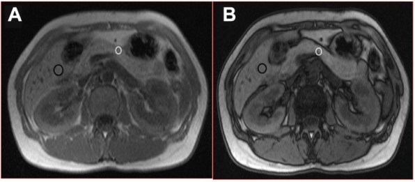
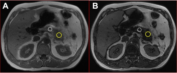
In this case control study, we retrospectively reviewed a local radiological and ERCP database to identify patients who had had abdominal magnetic resonance imaging (MRI) followed by ERCP no more than 60 days later between September 2003 and January 2011. Percentage of fat was determined by recording signal intensity in the in-phase (Sin) and out-of-phase (Sout) T1-weighted gradient sequences, and calculation of the fat fraction as (Sin - Sout)/(Sin) × 2 by an abdominal radiologist blinded to clinical history. Controls matched for age, gender, and other pancreatobiliary disease were selected from a group with no post-ERCP pancreatitis (before fat content of the pancreas was analyzed).

RESULTS

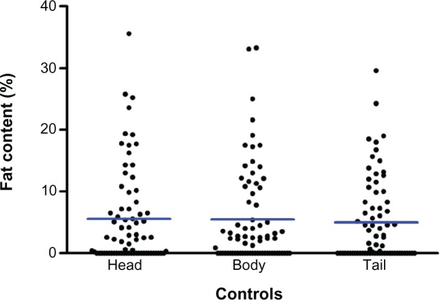
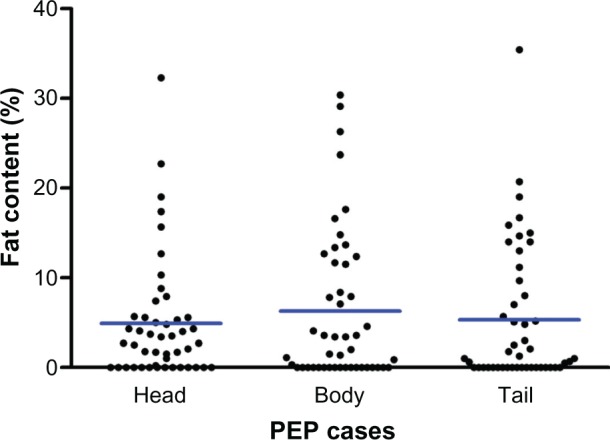
Forty-seven patients were enrolled. Compared with controls, subjects with post-ERCP pancreatitis were similar in terms of age (41.4 years versus 41.1 years), gender (21.2% versus 20.2% males), pancreatobiliary disease characteristics, and most ERCP techniques. Measurements of pancreatic head, body, and tail fat and body mass index were similar in patients and controls.

CONCLUSION

Increased pancreatic fat on MRI criteria is not an independent predictor of post-ERCP pancreatitis.

摘要

背景

一项初步研究显示,特发性胰腺炎和Oddi括约肌功能障碍患者的胰腺脂肪增加。在本研究中,我们旨在确定胰腺脂肪量增加是否是内镜逆行胰胆管造影术(ERCP)后胰腺炎的独立危险因素。

方法

在这项病例对照研究中,我们回顾性分析了本地放射学和ERCP数据库,以确定在2003年9月至2011年1月期间接受腹部磁共振成像(MRI)检查且在不超过60天后接受ERCP检查的患者。脂肪百分比通过记录同相位(Sin)和反相位(Sout)T1加权梯度序列中的信号强度来确定,并由对临床病史不知情的腹部放射科医生计算脂肪分数为(Sin - Sout)/(Sin)×2。从一组无ERCP后胰腺炎的患者(在分析胰腺脂肪含量之前)中选择年龄、性别和其他胰腺疾病相匹配的对照组。

结果

共纳入47例患者。与对照组相比,ERCP后胰腺炎患者在年龄(41.4岁对41.1岁)、性别(男性分别为21.2%对20.2%)、胰腺疾病特征以及大多数ERCP技术方面相似。患者和对照组在胰头、胰体、胰尾脂肪测量及体重指数方面相似。

结论

根据MRI标准,胰腺脂肪增加并非ERCP后胰腺炎的独立预测因素。

https://cdn.ncbi.nlm.nih.gov/pmc/blobs/0097/4061141/0a41a8b39cc6/ceg-7-199Fig4.jpg
https://cdn.ncbi.nlm.nih.gov/pmc/blobs/0097/4061141/177a647286c5/ceg-7-199Fig1.jpg
https://cdn.ncbi.nlm.nih.gov/pmc/blobs/0097/4061141/0bac1382e1a8/ceg-7-199Fig2.jpg
https://cdn.ncbi.nlm.nih.gov/pmc/blobs/0097/4061141/cf73531672a2/ceg-7-199Fig3.jpg
https://cdn.ncbi.nlm.nih.gov/pmc/blobs/0097/4061141/0a41a8b39cc6/ceg-7-199Fig4.jpg

相似文献

1
Increased fat in pancreas not associated with risk of pancreatitis post-endoscopic retrograde cholangiopancreatography.胰腺脂肪增加与内镜逆行胰胆管造影术后胰腺炎风险无关。
Clin Exp Gastroenterol. 2014 Jun 9;7:199-204. doi: 10.2147/CEG.S31333. eCollection 2014.
2
Outcomes of Endoscopic Retrograde Cholangiopancreatography (ERCP) and Sphincterotomy for Suspected Sphincter of Oddi Dysfunction (SOD) Post Roux-En-Y Gastric Bypass.内镜逆行胰胆管造影术(ERCP)及括约肌切开术治疗 Roux-en-Y 胃旁路术后疑似Oddi括约肌功能障碍(SOD)的疗效
Obes Surg. 2017 Oct;27(10):2656-2662. doi: 10.1007/s11695-017-2696-4.
3
Randomized controlled trial of pancreatic stenting to prevent pancreatitis after endoscopic retrograde cholangiopancreatography.随机对照试验:内镜逆行胰胆管造影术后胰管支架置入预防胰腺炎。
World J Gastroenterol. 2012 Apr 14;18(14):1635-41. doi: 10.3748/wjg.v18.i14.1635.
4
Risk factors for post-ERCP pancreatitis in high risk patients who have undergone prophylactic pancreatic duct stenting: a multicenter retrospective study.接受预防性胰管支架置入术的高危患者发生内镜逆行胰胆管造影术后胰腺炎的危险因素:一项多中心回顾性研究
Intern Med. 2011;50(24):2927-32. doi: 10.2169/internalmedicine.50.6235. Epub 2011 Dec 15.
5
Prophylactic pancreas stenting followed by needle-knife fistulotomy in patients with sphincter of Oddi dysfunction and difficult cannulation: new method to prevent post-ERCP pancreatitis.对Oddi括约肌功能障碍且插管困难的患者先进行预防性胰腺支架置入,然后行针刀瘘管切开术:预防内镜逆行胰胆管造影术后胰腺炎的新方法。
Dig Endosc. 2009 Jan;21(1):8-13. doi: 10.1111/j.1443-1661.2008.00819.x.
6
Post-endoscopic retrograde cholangiopancreatography pancreatitis: A systematic review for prevention and treatment.经内镜逆行胰胆管造影术后胰腺炎:预防和治疗的系统评价。
World J Gastroenterol. 2019 Aug 7;25(29):4019-4042. doi: 10.3748/wjg.v25.i29.4019.
7
Risk factors of post- endoscopic retrograde cholangiopancreatography pancreatitis in biliary type sphincter of Oddi dysfunction in Japanese patients.日本患者胆胰型 Oddi 括约肌功能障碍内镜逆行胰胆管造影术后胰腺炎的危险因素。
J Dig Dis. 2017 Oct;18(10):591-597. doi: 10.1111/1751-2980.12541.
8
Sphincter of Oddi manometry does not predispose to post-ERCP acute pancreatitis.Oddi括约肌测压不会增加ERCP术后急性胰腺炎的发病风险。
Gastrointest Endosc. 2004 Apr;59(4):499-505. doi: 10.1016/s0016-5107(03)02876-1.
9
The role of endoscopic retrograde cholangiopancreatography and endoscopic ultrasound in diagnosis and treatment of acute pancreatitis.内镜逆行胰胆管造影术和内镜超声在急性胰腺炎诊断与治疗中的作用。
Minerva Gastroenterol Dietol. 2005 Dec;51(4):265-88.
10
Pancreatic sphincter hypertension increases the risk of post-ERCP pancreatitis.胰括约肌高压会增加内镜逆行胰胆管造影术后胰腺炎的风险。
Endoscopy. 1997 May;29(4):252-7. doi: 10.1055/s-2007-1004185.

引用本文的文献

1
Evaluation of Development of Nonalcoholic Fatty Pancreas Disease After Post-Endoscopic Retrograde Cholangiop ancreatography Pancreatitis in Liver Transplant Patients: Computerized Tomography Versus Ultrasound.肝移植患者内镜逆行胰胆管造影术后胰腺炎后非酒精性脂肪性胰腺疾病发展的评估:计算机断层扫描与超声。
Turk J Gastroenterol. 2023 Nov;34(11):1180-1185. doi: 10.5152/tjg.2023.22424.
2
MRI assessment of ectopic fat accumulation in pancreas, liver and skeletal muscle in patients with obesity, overweight and normal BMI in correlation with the presence of central obesity and metabolic syndrome.肥胖、超重及体重指数正常的患者胰腺、肝脏和骨骼肌异位脂肪堆积的MRI评估及其与中心性肥胖和代谢综合征的相关性
Diabetes Metab Syndr Obes. 2019 May 3;12:623-636. doi: 10.2147/DMSO.S194690. eCollection 2019.
3

本文引用的文献

1
Noninvasive quantitation of human liver steatosis using magnetic resonance and bioassay methods.使用磁共振和生物测定方法对人类肝脏脂肪变性进行无创定量分析。
Eur Radiol. 2009 Aug;19(8):2033-40. doi: 10.1007/s00330-009-1351-4. Epub 2009 Mar 11.
2
Evaluation of diffuse liver steatosis by ultrasound, computed tomography, and magnetic resonance imaging: which modality is best?超声、计算机断层扫描和磁共振成像对弥漫性肝脂肪变性的评估:哪种方式最佳?
Clin Imaging. 2009 Mar-Apr;33(2):110-5. doi: 10.1016/j.clinimag.2008.06.036.
3
Chemical shift magnetic resonance imaging is helpful in detecting hepatic steatosis but not fibrosis in patients with nonalcoholic fatty liver disease (NAFLD).
Exploring the metabolic syndrome: Nonalcoholic fatty pancreas disease.探索代谢综合征:非酒精性脂肪性胰腺疾病。
World J Gastroenterol. 2016 Sep 14;22(34):7660-75. doi: 10.3748/wjg.v22.i34.7660.
4
Insulin Resistance as a Novel Risk Factor for Post-ERCP Pancreatitis: A Pilot Study.胰岛素抵抗作为内镜逆行胰胆管造影术后胰腺炎的一种新的危险因素:一项初步研究。
Dig Dis Sci. 2016 Aug;61(8):2397-2405. doi: 10.1007/s10620-016-4127-0. Epub 2016 Mar 19.
化学位移磁共振成像有助于检测非酒精性脂肪性肝病(NAFLD)患者的肝脂肪变性,但对肝纤维化检测无效。
Ann Hepatol. 2009 Jan-Mar;8(1):21-5.
4
Noninvasive assessment of hepatic steatosis.肝脂肪变性的无创评估。
Clin Gastroenterol Hepatol. 2009 Feb;7(2):135-40. doi: 10.1016/j.cgh.2008.11.023. Epub 2008 Dec 6.
5
Magnetic resonance imaging and spectroscopy for monitoring liver steatosis.用于监测肝脏脂肪变性的磁共振成像和波谱分析
J Magn Reson Imaging. 2008 Oct;28(4):937-45. doi: 10.1002/jmri.21542.
6
Liver steatosis: investigation of opposed-phase T1-weighted liver MR signal intensity loss and visceral fat measurement as biomarkers.肝脂肪变性:以反相位T1加权肝脏磁共振信号强度损失及内脏脂肪测量作为生物标志物的研究
Radiology. 2008 Oct;249(1):160-6. doi: 10.1148/radiol.2491071375.
7
A murine model of obesity implicates the adipokine milieu in the pathogenesis of severe acute pancreatitis.一种肥胖小鼠模型表明脂肪因子环境在重症急性胰腺炎的发病机制中起作用。
Am J Physiol Gastrointest Liver Physiol. 2008 Sep;295(3):G552-8. doi: 10.1152/ajpgi.90278.2008. Epub 2008 Jun 26.
8
Non-invasive means of measuring hepatic fat content.测量肝脏脂肪含量的非侵入性方法。
World J Gastroenterol. 2008 Jun 14;14(22):3476-83. doi: 10.3748/wjg.14.3476.
9
Nonalcoholic fatty pancreas disease.非酒精性脂肪胰腺病。
HPB (Oxford). 2007;9(4):312-8. doi: 10.1080/13651820701504157.
10
Hepato-pancreato-biliary fat: the good, the bad and the ugly.肝胆胰脂肪:好的、坏的和丑的。
HPB (Oxford). 2007;9(2):92-7. doi: 10.1080/13651820701286177.