Sundelin Karin, Petersen Anne, Soltanpour Yalda, Zetterberg Madeleine
Institute of Neuroscience and Physiology, Section of Clinical Neuroscience and Rehabilitation/Ophthalmology.
Institute of Biomedicine, Department of Medical Chemistry and Cell Biology, Sahlgrenska Academy at the University of Gothenburg, Sweden.
Open Ophthalmol J. 2014 May 30;8:19-23. doi: 10.2174/1874364101408010019. eCollection 2014.
Inter-individual differences in intrinsic proliferative capacity of lens epithelial cells may have importance for the risk of developing posterior capsule opacification (PCO) after cataract surgery. The purpose of the present study was to determine growth of human lens epithelial cells (HLEC) in culture and investigate possible associations with clinical characteristics of the donors, such as age, sex, pseudoexfoliation, uveitis and diabetes.
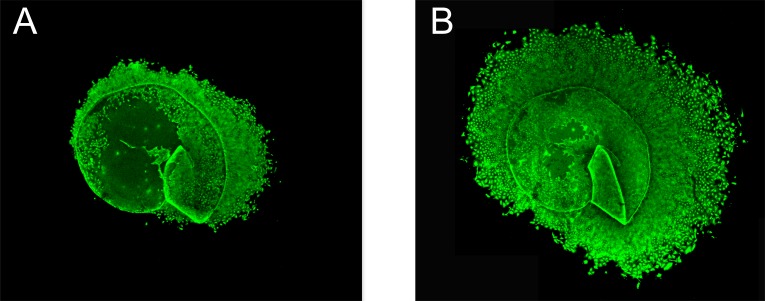
Pieces of lens capsule and adhering lens epithelial cells were obtained through capsulorhexis at cataract surgery. Specimens were cultured in a humidified CO2-incubator using standard culture medium and 5% fetal calf serum for two weeks after which cultured cells were stained with carboxy-fluorescein diacetate succinimidyl ester. Image processing software was used to determine the area of the confluent epithelial cell layer in relation to the size of the original capsule specimen.
The increase in area of confluent HLEC showed a negative correlation with diabetes at the first week after surgery. Lower age and female sex showed border-line significant associations with a higher rate of cell proliferation. The presence of pseudoexfoliation in vivo did not significantly affect cell growth in culture postoperatively. Nor did installation of xylocain in the anterior chamber during surgery.
Diabetes is associated with lower rate of proliferation of lens epithelial cells in culture. The lack of strong correlations between in vitro growth and known risk factors for PCO in the donors suggest that other factors than the proliferative capacity of the cells per se are important for PCO formation.
晶状体上皮细胞内在增殖能力的个体差异可能对白内障手术后发生后囊膜混浊(PCO)的风险具有重要意义。本研究的目的是确定培养的人晶状体上皮细胞(HLEC)的生长情况,并研究其与供体临床特征(如年龄、性别、假性剥脱、葡萄膜炎和糖尿病)之间可能存在的关联。
在白内障手术中通过撕囊获取晶状体囊膜碎片及附着的晶状体上皮细胞。标本在湿度可控的二氧化碳培养箱中,使用标准培养基和5%胎牛血清培养两周,之后用羧基荧光素二乙酸琥珀酰亚胺酯对培养细胞进行染色。使用图像处理软件确定汇合的上皮细胞层面积与原始囊膜标本大小的关系。
术后第一周,汇合的HLEC面积增加与糖尿病呈负相关。年龄较小和女性与较高的细胞增殖率呈边缘显著关联。体内存在假性剥脱对术后培养中的细胞生长无显著影响。手术期间在前房注射利多卡因也无显著影响。
糖尿病与培养的晶状体上皮细胞增殖率较低有关。体外生长与供体中已知的PCO风险因素之间缺乏强相关性,这表明除细胞本身的增殖能力外,其他因素对PCO的形成也很重要。