Parreno Justin, Emin Grace, Vu Michael P, Clark Jackson T, Aryal Sandeep, Patel Shaili D, Cheng Catherine
Department of Biological Sciences, University of Delaware, Newark, DE, United States.
Department of Biomedical Engineering, University of Delaware, Newark, DE, United States.
Front Cell Dev Biol. 2022 Sep 13;10:983178. doi: 10.3389/fcell.2022.983178. eCollection 2022.
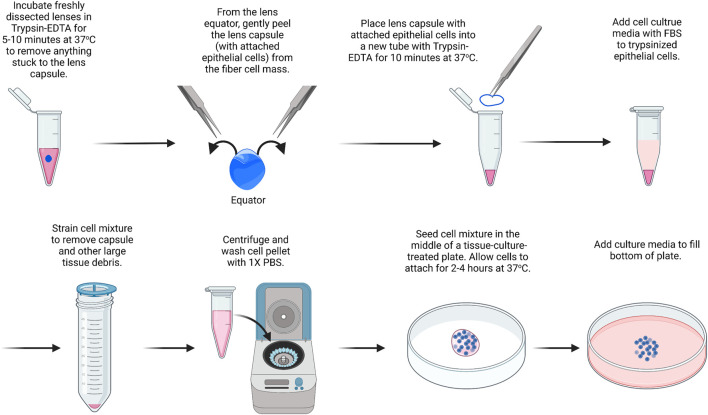
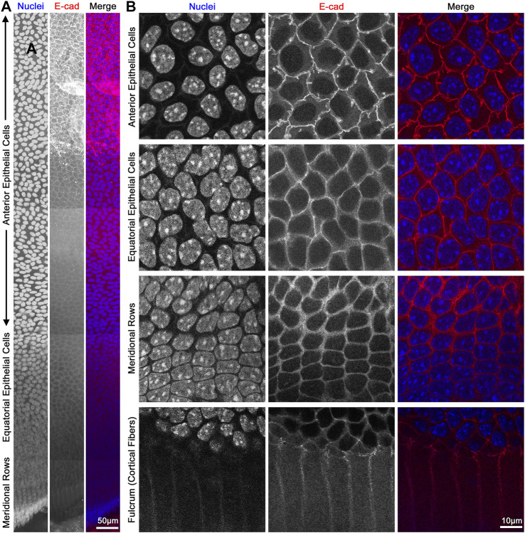
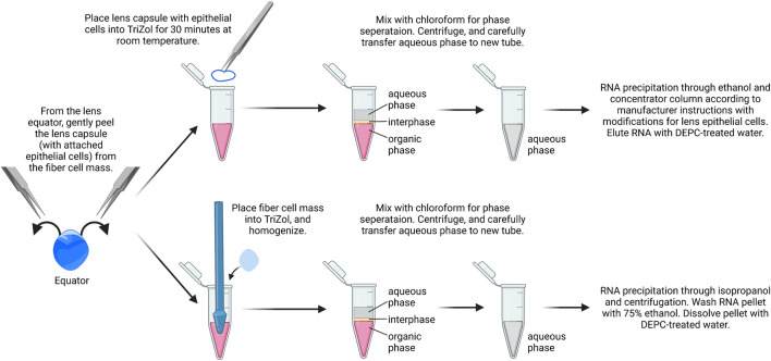
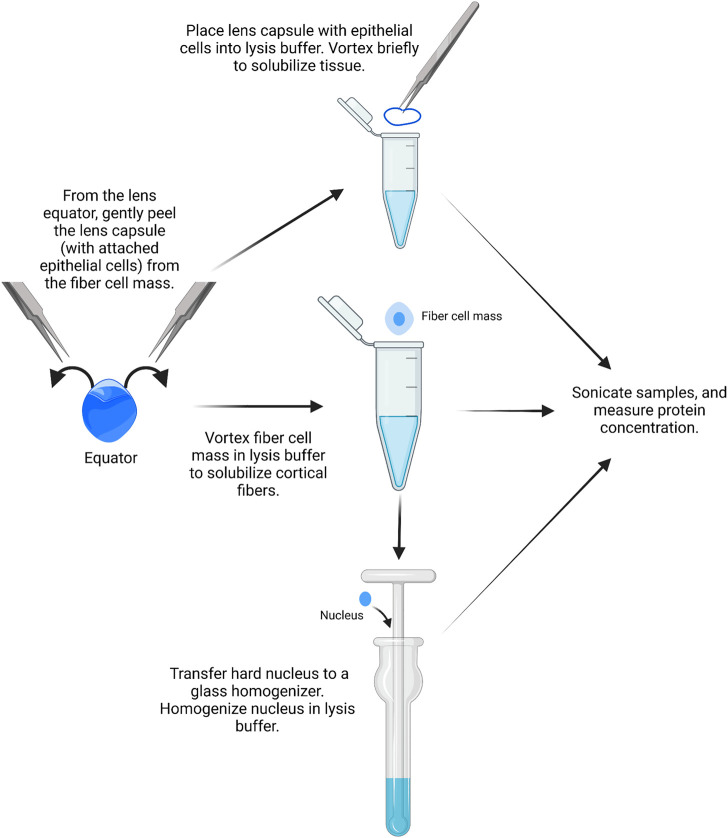
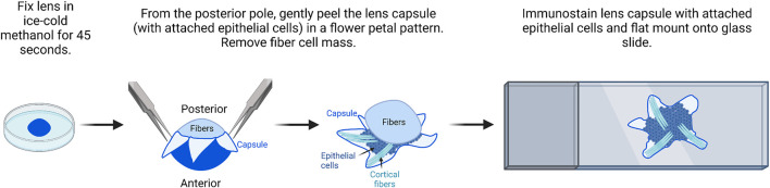
The transparent ocular lens in the anterior chamber of the eye is responsible for fine focusing of light onto the retina. The lens is entirely cellular with bulk of the tissue composed of fiber cells, and the anterior hemisphere of the lens is covered by a monolayer of epithelial cells. Lens epithelial cells are important for maintaining fiber cell homeostasis and for continual growth of the lens tissue throughout life. Cataracts, defined as any opacity in the lens, remain the leading cause of blindness in the world. Following cataract surgery, lens epithelial cells can undergo a process of epithelial-to-mesenchymal transition (EMT), leading to secondary cataracts due to posterior capsular opacification (PCO). Since the epithelial cells make up only a small fraction of the lens, specialized techniques are required to study lens epithelial cell biology and pathology. Studies using native lens epithelial cells often require pooling of samples to obtain enough cells to make sufficient samples for traditional molecular biology techniques. Here, we provide detailed protocols that enable the study of native mouse lens epithelial cells, including immunostaining of the native lens epithelium in flat mounts, extraction of RNA and proteins from pairs of lens epithelial monolayers, and isolation of lens epithelial cells for primary culture. These protocols will enable researchers to gain better insight on representative molecular expression and cellular structure of lens epithelial cells. We also provide comparative data between native, primary culture, and immortalized lens epithelial cells and discuss the advantages and disadvantages of each technique presented.
眼睛前房内的透明晶状体负责将光线精确聚焦到视网膜上。晶状体完全由细胞组成,其大部分组织由纤维细胞构成,晶状体的前半球被单层上皮细胞覆盖。晶状体上皮细胞对于维持纤维细胞的稳态以及晶状体组织在整个生命过程中的持续生长至关重要。白内障被定义为晶状体中的任何混浊,仍然是全球失明的主要原因。白内障手术后,晶状体上皮细胞可经历上皮 - 间充质转化(EMT)过程,由于后囊膜混浊(PCO)导致继发性白内障。由于上皮细胞仅占晶状体的一小部分,因此需要专门的技术来研究晶状体上皮细胞生物学和病理学。使用天然晶状体上皮细胞的研究通常需要汇集样本以获得足够的细胞,从而为传统分子生物学技术制备足够的样本。在这里,我们提供了详细的方案,用于研究天然小鼠晶状体上皮细胞,包括在扁平标本上对天然晶状体上皮进行免疫染色、从成对的晶状体上皮单层中提取RNA和蛋白质,以及分离晶状体上皮细胞进行原代培养。这些方案将使研究人员能够更好地了解晶状体上皮细胞的代表性分子表达和细胞结构。我们还提供了天然、原代培养和永生化晶状体上皮细胞之间的比较数据,并讨论了所介绍的每种技术的优缺点。