• 文献检索
  • 文档翻译
  • 深度研究
  • 学术资讯
  • Suppr Zotero 插件Zotero 插件
  • 邀请有礼
  • 套餐&价格
  • 历史记录
应用&插件
Suppr Zotero 插件Zotero 插件浏览器插件Mac 客户端Windows 客户端微信小程序
定价
高级版会员购买积分包购买API积分包
服务
文献检索文档翻译深度研究API 文档MCP 服务
关于我们
关于 Suppr公司介绍联系我们用户协议隐私条款
关注我们

Suppr 超能文献

核心技术专利:CN118964589B侵权必究
粤ICP备2023148730 号-1Suppr @ 2026

文献检索

告别复杂PubMed语法,用中文像聊天一样搜索,搜遍4000万医学文献。AI智能推荐,让科研检索更轻松。

立即免费搜索

文件翻译

保留排版,准确专业,支持PDF/Word/PPT等文件格式,支持 12+语言互译。

免费翻译文档

深度研究

AI帮你快速写综述,25分钟生成高质量综述,智能提取关键信息,辅助科研写作。

立即免费体验

SHERPA:用于硅藻和其他物体的图像分割和轮廓特征提取工具。

SHERPA: an image segmentation and outline feature extraction tool for diatoms and other objects.

机构信息

AWI: Alfred Wegener Institute, Helmholtz Centre for Polar and Marine Research, Am Handelshafen 12, 27570 Bremerhaven, Germany.

出版信息

BMC Bioinformatics. 2014 Jun 25;15:218. doi: 10.1186/1471-2105-15-218.

DOI:10.1186/1471-2105-15-218
PMID:24964954
原文链接:https://pmc.ncbi.nlm.nih.gov/articles/PMC4087246/
Abstract

BACKGROUND

Light microscopic analysis of diatom frustules is widely used both in basic and applied research, notably taxonomy, morphometrics, water quality monitoring and paleo-environmental studies. In these applications, usually large numbers of frustules need to be identified and/or measured. Although there is a need for automation in these applications, and image processing and analysis methods supporting these tasks have previously been developed, they did not become widespread in diatom analysis. While methodological reports for a wide variety of methods for image segmentation, diatom identification and feature extraction are available, no single implementation combining a subset of these into a readily applicable workflow accessible to diatomists exists.

RESULTS

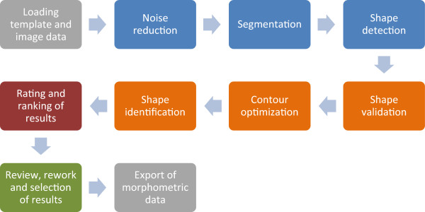
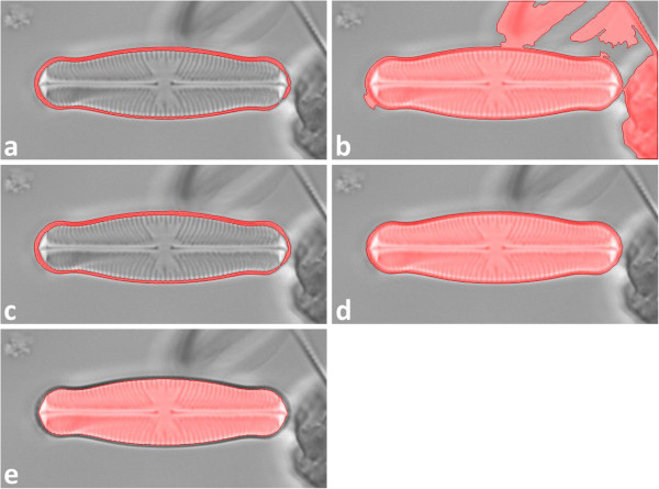
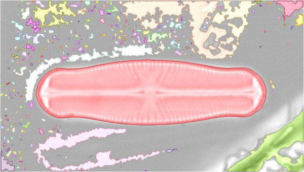
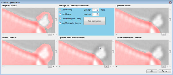
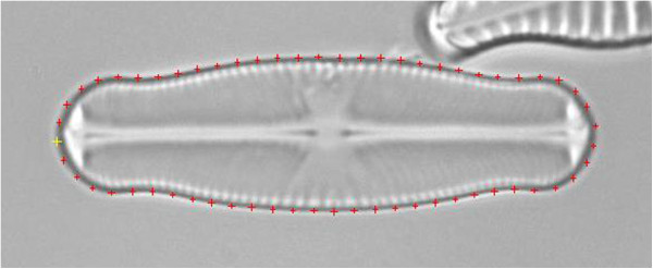
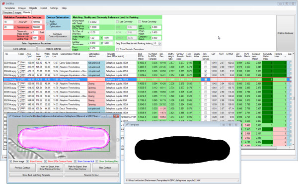
The newly developed tool SHERPA offers a versatile image processing workflow focused on the identification and measurement of object outlines, handling all steps from image segmentation over object identification to feature extraction, and providing interactive functions for reviewing and revising results. Special attention was given to ease of use, applicability to a broad range of data and problems, and supporting high throughput analyses with minimal manual intervention.

CONCLUSIONS

Tested with several diatom datasets from different sources and of various compositions, SHERPA proved its ability to successfully analyze large amounts of diatom micrographs depicting a broad range of species. SHERPA is unique in combining the following features: application of multiple segmentation methods and selection of the one giving the best result for each individual object; identification of shapes of interest based on outline matching against a template library; quality scoring and ranking of resulting outlines supporting quick quality checking; extraction of a wide range of outline shape descriptors widely used in diatom studies and elsewhere; minimizing the need for, but enabling manual quality control and corrections. Although primarily developed for analyzing images of diatom valves originating from automated microscopy, SHERPA can also be useful for other object detection, segmentation and outline-based identification problems.

摘要

背景

基于对硅藻类化石壳的微观分析,在基础和应用研究领域都有着广泛的应用,尤其在分类学、形态计量学、水质监测和古环境研究等方面。在这些应用中,通常需要识别和/或测量大量的化石壳。尽管这些应用需要自动化,并且已经开发了支持这些任务的图像处理和分析方法,但它们在硅藻类化石分析中并没有得到广泛应用。虽然有各种用于图像分割、硅藻类鉴定和特征提取的方法的方法报告,但没有一个单一的实现方案将这些方法中的一部分组合成一个易于应用的、可供硅藻类化石学家使用的工作流程。

结果

新开发的工具 SHERPA 提供了一个通用的图像处理工作流程,重点是识别和测量物体轮廓,处理从图像分割到物体识别再到特征提取的所有步骤,并提供交互功能,用于检查和修改结果。特别注意易用性、适用于广泛的数据和问题,以及支持最小人工干预的高通量分析。

结论

通过对来自不同来源和不同组成的几个硅藻类数据集进行测试,SHERPA 证明了它能够成功地分析大量描绘广泛物种的硅藻类显微照片。SHERPA 的独特之处在于结合了以下功能:应用多种分割方法,并为每个单独的物体选择最佳的分割结果;基于与模板库的轮廓匹配来识别感兴趣的形状;对结果轮廓进行质量评分和排名,支持快速质量检查;提取广泛用于硅藻类研究和其他领域的轮廓形状描述符;最大限度地减少手动质量控制和修正的需求,但同时也允许进行。虽然 SHERPA 主要是为分析来自自动化显微镜的硅藻类化石壳图像而开发的,但它也可用于其他物体检测、分割和基于轮廓的识别问题。

https://cdn.ncbi.nlm.nih.gov/pmc/blobs/8612/4087246/80adc284404c/1471-2105-15-218-15.jpg
https://cdn.ncbi.nlm.nih.gov/pmc/blobs/8612/4087246/0f4471e69202/1471-2105-15-218-1.jpg
https://cdn.ncbi.nlm.nih.gov/pmc/blobs/8612/4087246/775faab62177/1471-2105-15-218-2.jpg
https://cdn.ncbi.nlm.nih.gov/pmc/blobs/8612/4087246/ad3df18f9b96/1471-2105-15-218-3.jpg
https://cdn.ncbi.nlm.nih.gov/pmc/blobs/8612/4087246/a0781216ac34/1471-2105-15-218-4.jpg
https://cdn.ncbi.nlm.nih.gov/pmc/blobs/8612/4087246/7238832c2d3b/1471-2105-15-218-5.jpg
https://cdn.ncbi.nlm.nih.gov/pmc/blobs/8612/4087246/0c261a058048/1471-2105-15-218-6.jpg
https://cdn.ncbi.nlm.nih.gov/pmc/blobs/8612/4087246/7392e143c54c/1471-2105-15-218-7.jpg
https://cdn.ncbi.nlm.nih.gov/pmc/blobs/8612/4087246/10b5d5547246/1471-2105-15-218-8.jpg
https://cdn.ncbi.nlm.nih.gov/pmc/blobs/8612/4087246/8a6bb9655070/1471-2105-15-218-9.jpg
https://cdn.ncbi.nlm.nih.gov/pmc/blobs/8612/4087246/bae7cc7b8afc/1471-2105-15-218-10.jpg
https://cdn.ncbi.nlm.nih.gov/pmc/blobs/8612/4087246/a2a9a205195e/1471-2105-15-218-11.jpg
https://cdn.ncbi.nlm.nih.gov/pmc/blobs/8612/4087246/f348bc0f6bd7/1471-2105-15-218-12.jpg
https://cdn.ncbi.nlm.nih.gov/pmc/blobs/8612/4087246/a311f193ba31/1471-2105-15-218-13.jpg
https://cdn.ncbi.nlm.nih.gov/pmc/blobs/8612/4087246/eeb3f9b2aa4d/1471-2105-15-218-14.jpg
https://cdn.ncbi.nlm.nih.gov/pmc/blobs/8612/4087246/80adc284404c/1471-2105-15-218-15.jpg

相似文献

1
SHERPA: an image segmentation and outline feature extraction tool for diatoms and other objects.SHERPA:用于硅藻和其他物体的图像分割和轮廓特征提取工具。
BMC Bioinformatics. 2014 Jun 25;15:218. doi: 10.1186/1471-2105-15-218.
2
Improving deep learning-based segmentation of diatoms in gigapixel-sized virtual slides by object-based tile positioning and object integrity constraint.通过基于对象的图块定位和对象完整性约束来改进千兆像素大小虚拟幻灯片中基于深度学习的硅藻分割。
PLoS One. 2023 Feb 24;18(2):e0272103. doi: 10.1371/journal.pone.0272103. eCollection 2023.
3
Multi-template matching: a versatile tool for object-localization in microscopy images.多模板匹配:显微镜图像中目标定位的通用工具。
BMC Bioinformatics. 2020 Feb 5;21(1):44. doi: 10.1186/s12859-020-3363-7.
4
Quantitative comparison of taxa and taxon concepts in the diatom genus Fragilariopsis: a case study on using slide scanning, multiexpert image annotation, and image analysis in taxonomy.定量比较硅藻属 Fragilariopsis 中的分类单元和分类概念:使用幻灯片扫描、多专家图像注释和图像分析进行分类学研究的案例。
J Phycol. 2018 Oct;54(5):703-719. doi: 10.1111/jpy.12767. Epub 2018 Aug 28.
5
Automatic segmentation of diatom images for classification.用于分类的硅藻图像自动分割
Microsc Res Tech. 2004 Sep;65(1-2):72-85. doi: 10.1002/jemt.20111.
6
A preliminary study on automated freshwater algae recognition and classification system.自动化淡水藻类识别与分类系统初探
BMC Bioinformatics. 2012;13 Suppl 17(Suppl 17):S25. doi: 10.1186/1471-2105-13-S17-S25. Epub 2012 Dec 13.
7
Metabarcoding vs. morphological identification to assess diatom diversity in environmental studies.代谢条码与形态鉴定在环境研究中评估硅藻多样性的比较。
Mol Ecol Resour. 2015 May;15(3):526-42. doi: 10.1111/1755-0998.12336. Epub 2014 Oct 20.
8
Polymorph segmentation representation for medical image computing.多态分割表示在医学图像计算中的应用。
Comput Methods Programs Biomed. 2019 Apr;171:19-26. doi: 10.1016/j.cmpb.2019.02.011. Epub 2019 Feb 21.
9
From image to data using common image-processing techniques.运用常见图像处理技术从图像获取数据。
Curr Protoc Cytom. 2010 Oct;Chapter 12:Unit 12.21. doi: 10.1002/0471142956.cy1221s54.
10
Automated diatom searching in the digital scanning electron microscopy images of drowning cases using the deep neural networks.利用深度神经网络对溺亡案件的数字扫描电子显微镜图像进行自动化硅藻搜索。
Int J Legal Med. 2021 Mar;135(2):497-508. doi: 10.1007/s00414-020-02392-z. Epub 2020 Aug 13.

引用本文的文献

1
GRABSEEDS: extraction of plant organ traits through image analysis.GRABSEEDS:通过图像分析提取植物器官特征
Plant Methods. 2024 Sep 12;20(1):140. doi: 10.1186/s13007-024-01268-2.
2
A lightweight convolutional neural network (CNN) model for diatom classification: DiatomNet.一种用于硅藻分类的轻量级卷积神经网络(CNN)模型:硅藻网络。
PeerJ Comput Sci. 2024 Mar 29;10:e1970. doi: 10.7717/peerj-cs.1970. eCollection 2024.
3
Improving deep learning-based segmentation of diatoms in gigapixel-sized virtual slides by object-based tile positioning and object integrity constraint.

本文引用的文献

1
Does the cosmopolitan diatom Gomphonema parvulum (Kützing) Kützing have a biogeography?世界性硅藻小异极藻(库津)库津是否具有生物地理学特征?
PLoS One. 2014 Jan 29;9(1):e86885. doi: 10.1371/journal.pone.0086885. eCollection 2014.
2
First evidence of the existence of semi-cryptic species and of a phylogeographic structure in the Gomphonema parvulum (Kützing) Kützing complex (Bacillariophyta).首次发现颗粒直链藻(Kützing)复合体(Bacillariophyta)中半隐种的存在和系统地理结构的证据。
Protist. 2013 Sep;164(5):686-705. doi: 10.1016/j.protis.2013.07.005. Epub 2013 Aug 28.
3
An inordinate fondness? The number, distributions, and origins of diatom species.
通过基于对象的图块定位和对象完整性约束来改进千兆像素大小虚拟幻灯片中基于深度学习的硅藻分割。
PLoS One. 2023 Feb 24;18(2):e0272103. doi: 10.1371/journal.pone.0272103. eCollection 2023.
4
Phylotranscriptomics reveals the reticulate evolutionary history of a widespread diatom species complex.系统发生转录组学揭示了一种广泛分布的硅藻种复合体的网状进化历史。
J Phycol. 2022 Oct;58(5):643-656. doi: 10.1111/jpy.13281. Epub 2022 Aug 8.
5
SizeExtractR: A workflow for rapid reproducible extraction of object size metrics from scaled images.SizeExtractR:一种从缩放图像中快速可重复提取对象大小度量的工作流程。
Ecol Evol. 2022 Mar 14;12(3):e8724. doi: 10.1002/ece3.8724. eCollection 2022 Mar.
6
Repositories for Taxonomic Data: Where We Are and What is Missing.分类学数据存储库:我们的现状和缺失的内容。
Syst Biol. 2020 Nov 1;69(6):1231-1253. doi: 10.1093/sysbio/syaa026.
7
A Patented Rapid Method for Identification of Italian Diatom Species.一种已获专利的快速鉴定意大利硅藻物种的方法。
Int J Environ Res Public Health. 2019 Oct 16;16(20):3933. doi: 10.3390/ijerph16203933.
8
Diatom identification including life cycle stages through morphological and texture descriptors.通过形态和纹理描述符进行硅藻鉴定,包括生命周期阶段。
PeerJ. 2019 Apr 25;7:e6770. doi: 10.7717/peerj.6770. eCollection 2019.
9
Shape outline extraction software (DiaOutline) for elliptic Fourier analysis application in morphometric studies.用于形态计量学研究中椭圆傅里叶分析的形状轮廓提取软件(DiaOutline)。
Appl Plant Sci. 2018 Dec 21;6(12):e01204. doi: 10.1002/aps3.1204. eCollection 2018 Dec.
10
Quantitative comparison of taxa and taxon concepts in the diatom genus Fragilariopsis: a case study on using slide scanning, multiexpert image annotation, and image analysis in taxonomy.定量比较硅藻属 Fragilariopsis 中的分类单元和分类概念:使用幻灯片扫描、多专家图像注释和图像分析进行分类学研究的案例。
J Phycol. 2018 Oct;54(5):703-719. doi: 10.1111/jpy.12767. Epub 2018 Aug 28.
过分的喜爱?硅藻物种的数量、分布和起源。
J Eukaryot Microbiol. 2013 Jul-Aug;60(4):414-20. doi: 10.1111/jeu.12047. Epub 2013 May 27.
4
A computational approach to edge detection.一种基于计算的边缘检测方法。
IEEE Trans Pattern Anal Mach Intell. 1986 Jun;8(6):679-98.
5
MorphoJ: an integrated software package for geometric morphometrics.MorphoJ:一个用于几何形态测量学的集成软件包。
Mol Ecol Resour. 2011 Mar;11(2):353-7. doi: 10.1111/j.1755-0998.2010.02924.x. Epub 2010 Oct 5.
6
Pseudocryptic diversity versus cosmopolitanism in diatoms: a case study on Navicula cryptocephala Kütz. (Bacillariophyceae) and morphologically similar taxa.拟隐种多样性与硅藻的世界性:以 Navicula cryptocephala Kütz.(Bacillariophyceae)和形态相似分类群为例。
Protist. 2010 Jul;161(3):353-69. doi: 10.1016/j.protis.2009.12.003. Epub 2010 Jan 22.
7
Diatom elemental and morphological changes in response to iron limitation: a brief review with potential paleoceanographic applications.硅藻对铁限制的元素和形态变化:具有潜在古海洋学应用的简要综述
Geobiology. 2009 Sep;7(4):419-31. doi: 10.1111/j.1472-4669.2009.00207.x. Epub 2009 Jul 29.
8
Improvement of a retinal blood vessel segmentation method using the Insight Segmentation and Registration Toolkit (ITK).使用洞察分割与配准工具包(ITK)改进视网膜血管分割方法。
Annu Int Conf IEEE Eng Med Biol Soc. 2007;2007:892-5. doi: 10.1109/IEMBS.2007.4352434.