Fayad Laura M, Blakeley Jaishri, Plotkin Scott, Widemann Brigitte, Jacobs Michael A
The Russell H. Morgan Department of Radiology and Radiological Science, The Johns Hopkins University School of Medicine, Baltimore, MD 21287, USA ; Sidney Kimmel Comprehensive Cancer Center, The Johns Hopkins University School of Medicine, Baltimore, MD 21287, USA ; Department of Orthopaedic Surgery, The Johns Hopkins University School of Medicine, Baltimore, MD 21287, USA.
Sidney Kimmel Comprehensive Cancer Center, The Johns Hopkins University School of Medicine, Baltimore, MD 21287, USA ; The Johns Hopkins Hospital Comprehensive Neurofibromatosis Center, Department of Neurology, The Johns Hopkins Hospital, CRB II, Suite 1M16, 1550 Orleans Street, Baltimore, MD 21231, USA.
ISRN Radiol. 2013 Oct 7;2013:627932. doi: 10.5402/2013/627932. eCollection 2013.
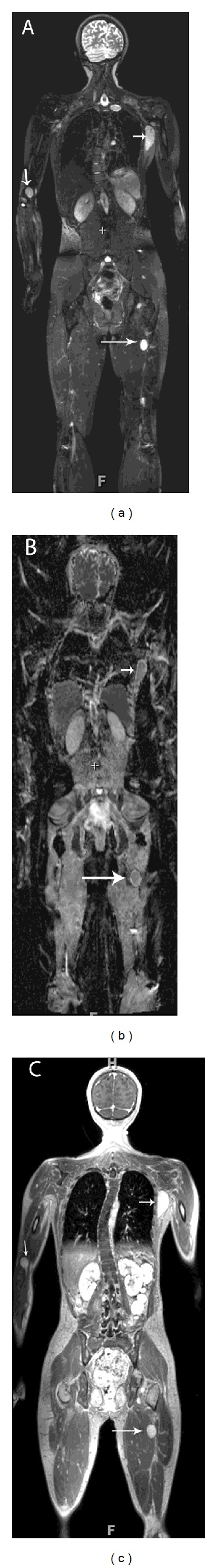
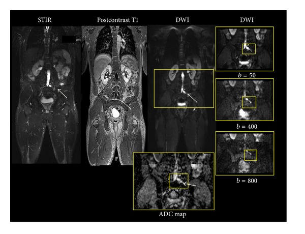
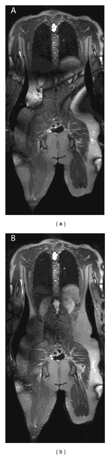
Purpose. WB-MRI is mainly used for tumor detection and surveillance. The purpose of this study is to establish the feasibility of WB-MRI at 3T for lesion characterization, with DWI/ADC-mapping and contrast-enhanced sequences, in patients with neurofibromatosis type 2 (NF-2) and schwannomatosis. Materials and Methods. At 3T, WB-MRI was performed in 11 subjects (10 NF-2 and 1 schwannomatosis) with STIR, T1, contrast-enhanced T1, and DWI/ADC mapping (b = 50, 400, 800 s/mm(2)). Two readers reviewed imaging for the presence and character of peripheral lesions. Lesion size and features (signal intensity, heterogeneity, enhancement characteristics, and ADC values) were recorded. Descriptive statistics were reported. Results. Twenty-three lesions were identified, with average size of 4.6 ± 2.8 cm. Lesions were characterized as tumors (21/23) or cysts (2/23) by contrast-enhancement properties (enhancement in tumors, no enhancement in cysts). On T1, tumors were homogeneously isointense (5/21) or hypointense (16/21); on STIR, tumors were hyperintense and homogeneous (10/21) or heterogeneous (11/21); on postcontrast T1, tumors enhanced homogeneously (14/21) or heterogeneously (7/21); on DWI, tumor ADC values were variable (range 0.8-2.7), suggesting variability in intrinsic tumor properties. Conclusion. WB-MRI with quantitative DWI and contrast-enhanced sequences at 3T is feasible and advances the utility of WB-MRI not only to include detection, but also to provide additional metrics for lesion characterization.
目的。全身弥散加权磁共振成像(WB-MRI)主要用于肿瘤检测和监测。本研究的目的是确定在3T场强下,利用弥散加权成像/表观扩散系数(DWI/ADC)映射和对比增强序列对2型神经纤维瘤病(NF-2)和神经鞘瘤病患者的病变进行特征性描述时,WB-MRI的可行性。材料与方法。在3T场强下,对11名受试者(10名NF-2患者和1名神经鞘瘤病患者)进行WB-MRI检查,扫描序列包括短TI反转恢复(STIR)序列、T1加权像、对比增强T1加权像以及DWI/ADC映射(b值 = 50、400、800 s/mm²)。两名阅片者对周围病变的存在情况和特征进行影像学评估。记录病变大小和特征(信号强度、异质性、增强特征和ADC值)。报告描述性统计结果。结果。共识别出23个病变,平均大小为4.6 ± 2.8 cm。根据对比增强特性(肿瘤有增强,囊肿无增强),病变被分为肿瘤(21/23)或囊肿(2/23)。在T1加权像上,肿瘤呈均匀等信号(5/21)或低信号(16/21);在STIR序列上,肿瘤呈高信号,均匀(10/21)或不均匀(11/21);在对比增强T1加权像上,肿瘤呈均匀增强(14/21)或不均匀增强(7/21);在DWI上,肿瘤的ADC值各不相同(范围为0.8 - 2.7),提示肿瘤内在特性存在差异。结论。3T场强下采用定量DWI和对比增强序列的WB-MRI是可行的,不仅提高了WB-MRI在肿瘤检测方面的应用,还为病变特征性描述提供了更多指标。