• 文献检索
  • 文档翻译
  • 深度研究
  • 学术资讯
  • Suppr Zotero 插件Zotero 插件
  • 邀请有礼
  • 套餐&价格
  • 历史记录
应用&插件
Suppr Zotero 插件Zotero 插件浏览器插件Mac 客户端Windows 客户端微信小程序
定价
高级版会员购买积分包购买API积分包
服务
文献检索文档翻译深度研究API 文档MCP 服务
关于我们
关于 Suppr公司介绍联系我们用户协议隐私条款
关注我们

Suppr 超能文献

核心技术专利:CN118964589B侵权必究
粤ICP备2023148730 号-1Suppr @ 2026

文献检索

告别复杂PubMed语法,用中文像聊天一样搜索,搜遍4000万医学文献。AI智能推荐,让科研检索更轻松。

立即免费搜索

文件翻译

保留排版,准确专业,支持PDF/Word/PPT等文件格式,支持 12+语言互译。

免费翻译文档

深度研究

AI帮你快速写综述,25分钟生成高质量综述,智能提取关键信息,辅助科研写作。

立即免费体验

多光子成像技术用于鉴定完整叶片中的类囊体、基质类囊体和淀粉粒。

Multiphoton imaging to identify grana, stroma thylakoid, and starch inside an intact leaf.

机构信息

Department of Physics, National Taiwan University, Taipei, Taiwan.

出版信息

BMC Plant Biol. 2014 Jun 27;14:175. doi: 10.1186/1471-2229-14-175.

DOI:10.1186/1471-2229-14-175
PMID:24969621
原文链接:https://pmc.ncbi.nlm.nih.gov/articles/PMC4104400/
Abstract

BACKGROUND

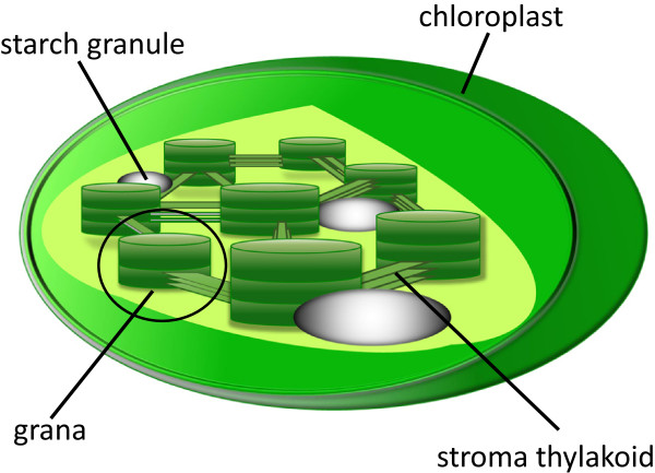
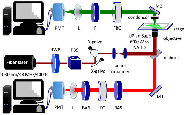
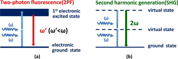
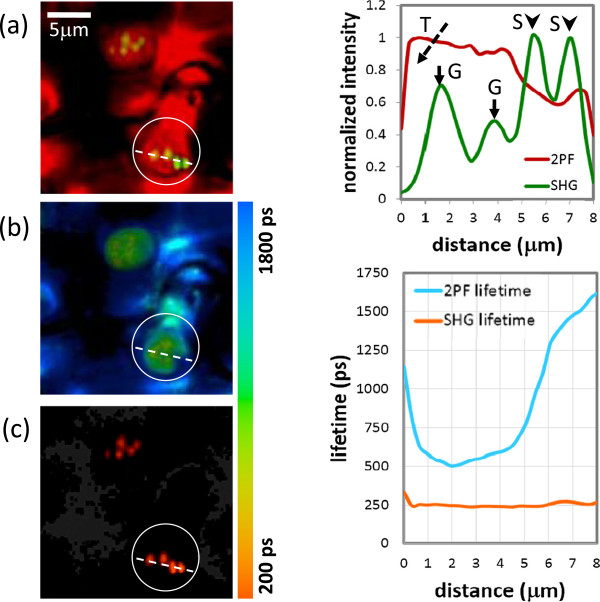
Grana and starch are major functional structures for photosynthesis and energy storage of plant, respectively. Both exhibit highly ordered molecular structures and appear as micrometer-sized granules inside chloroplasts. In order to distinguish grana and starch, we used multiphoton microscopy, with simultaneous acquisition of two-photon fluorescence (2PF) and second harmonic generation (SHG) signals. SHG is sensitive to crystallized structures while 2PF selectively reveals the distribution of chlorophyll.

RESULT

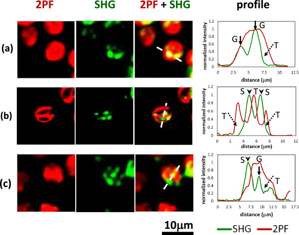
Three distinct microstructures with different contrasts were observed, i.e. "SHG dominates", "2PF dominates", and "SHG collocated with 2PF". It is known that starch and grana both emit SHG due to their highly crystallized structures, and no autofluorescence is emitted from starch, so the "SHG dominates" contrast should correspond to starch. The contrast of "SHG collocated with 2PF" is assigned to be grana, which exhibit crystallized structure with autofluorescent chlorophyll. The "2PF dominates" contrast should correspond to stroma thylakoid, which is a non-packed membrane structure with chrolophyll. The contrast assignment is further supported by fluorescence lifetime measurement.

CONCLUSION

We have demonstrated a straightforward and noninvasive method to identify the distribution of grana and starch within an intact leaf. By merging the 2PF and SHG images, grana, starch and stroma thylakoid can be visually distinguished. This approach can be extended to the observation of 3D grana distribution and their dynamics in living plants.

摘要

背景

类囊体基粒和淀粉粒分别是植物光合作用和能量储存的主要功能结构。它们都具有高度有序的分子结构,在叶绿体中呈现为微米级颗粒。为了区分基粒和淀粉粒,我们使用多光子显微镜,同时获取双光子荧光(2PF)和二次谐波产生(SHG)信号。SHG 对结晶结构敏感,而 2PF 则选择性地揭示叶绿素的分布。

结果

观察到三种具有不同对比度的不同微观结构,即“SHG 占主导”、“2PF 占主导”和“SHG 与 2PF 共定位”。已知淀粉粒和基粒都由于其高度结晶的结构而发出 SHG,并且淀粉粒不发出自发荧光,因此“SHG 占主导”的对比度应该对应于淀粉粒。“SHG 与 2PF 共定位”的对比度被分配给基粒,基粒具有结晶结构和自体荧光叶绿素。“2PF 占主导”的对比度应该对应于基质类囊体,它是一种具有叶绿素的非堆积膜结构。荧光寿命测量进一步支持了对比度的分配。

结论

我们已经证明了一种简单、非侵入性的方法,可以识别完整叶片中基粒和淀粉粒的分布。通过合并 2PF 和 SHG 图像,可以直观地区分基粒、淀粉粒和基质类囊体。这种方法可以扩展到观察活植物中 3D 基粒分布及其动态。

https://cdn.ncbi.nlm.nih.gov/pmc/blobs/cf62/4104400/32a22322bf13/1471-2229-14-175-5.jpg
https://cdn.ncbi.nlm.nih.gov/pmc/blobs/cf62/4104400/68c7eca7b5c8/1471-2229-14-175-1.jpg
https://cdn.ncbi.nlm.nih.gov/pmc/blobs/cf62/4104400/e31a3a054454/1471-2229-14-175-2.jpg
https://cdn.ncbi.nlm.nih.gov/pmc/blobs/cf62/4104400/3de25625a3a8/1471-2229-14-175-3.jpg
https://cdn.ncbi.nlm.nih.gov/pmc/blobs/cf62/4104400/d397e4304561/1471-2229-14-175-4.jpg
https://cdn.ncbi.nlm.nih.gov/pmc/blobs/cf62/4104400/32a22322bf13/1471-2229-14-175-5.jpg

相似文献

1
Multiphoton imaging to identify grana, stroma thylakoid, and starch inside an intact leaf.多光子成像技术用于鉴定完整叶片中的类囊体、基质类囊体和淀粉粒。
BMC Plant Biol. 2014 Jun 27;14:175. doi: 10.1186/1471-2229-14-175.
2
Second harmonic imaging of chloroplasts using the two-photon laser scanning microscope.使用双光子激光扫描显微镜对叶绿体进行二次谐波成像。
Micron. 2009 Apr;40(3):378-85. doi: 10.1016/j.micron.2008.09.007. Epub 2008 Oct 15.
3
Size-Dependent Optical Properties of Grana Inside Chloroplast of Plant Cells.植物细胞叶绿体类囊体内大小依赖的光学性质。
J Phys Chem B. 2017 Feb 9;121(5):915-922. doi: 10.1021/acs.jpcb.6b10204. Epub 2017 Jan 26.
4
Significance of structural variation in thylakoid membranes in maintaining functional photosystems during reproductive growth.类囊体膜结构变异在生殖生长期间维持功能性光系统中的意义。
Physiol Plant. 2017 May;160(1):111-123. doi: 10.1111/ppl.12528. Epub 2016 Dec 28.
5
The Reason for Growth Inhibition of 'Jinye': Lower Resistance and Abnormal Development of Chloroplasts Slow Down the Accumulation of Energy.“金叶”生长抑制的原因:较低的抗性和叶绿体的异常发育减缓了能量的积累。
Int J Mol Sci. 2019 Aug 29;20(17):4227. doi: 10.3390/ijms20174227.
6
Changes in chloroplast ultrastructure, fatty acid components of thylakoid membrane and chlorophyll a fluorescence transient in flag leaves of a super-high-yield hybrid rice and its parents during the reproductive stage.在生殖阶段,超高产杂交水稻及其亲本的叶绿体超微结构、类囊体膜脂肪酸成分和叶绿素 a 荧光动力学的变化。
J Plant Physiol. 2010 Mar 1;167(4):277-85. doi: 10.1016/j.jplph.2009.09.017. Epub 2009 Dec 9.
7
Three-dimensional architecture of grana and stroma thylakoids of higher plants as determined by electron tomography.高等植物类囊体垛叠和基质类囊体的三维结构通过电子断层扫描技术确定。
Plant Physiol. 2011 Apr;155(4):1601-11. doi: 10.1104/pp.110.170647. Epub 2011 Jan 11.
8
Arabidopsis CURVATURE THYLAKOID1 proteins modify thylakoid architecture by inducing membrane curvature.拟南芥 CURVATURE THYLAKOID1 蛋白通过诱导膜曲率来改变类囊体结构。
Plant Cell. 2013 Jul;25(7):2661-78. doi: 10.1105/tpc.113.113118. Epub 2013 Jul 9.
9
Fractionation of thylakoid membranes into grana and stroma thylakoids.将类囊体膜分离成基粒类囊体和基质类囊体。
Methods Mol Biol. 2004;274:1-9. doi: 10.1385/1-59259-799-8:001.
10
Analysis of the polypeptide composition of grana and stroma thylakoids by two-dimensional gel electrophoresis. Distribution of photosystem II between grana and stroma lamellae.通过二维凝胶电泳分析基粒和基质类囊体的多肽组成。光系统II在基粒和基质片层之间的分布。
Eur J Biochem. 1987 Dec 1;169(2):283-8. doi: 10.1111/j.1432-1033.1987.tb13609.x.

引用本文的文献

1
Simultaneous 3D Construction and Imaging of Plant Cells Using Plasmonic Nanoprobe-Assisted Multimodal Nonlinear Optical Microscopy.使用等离子体纳米探针辅助多模态非线性光学显微镜对植物细胞进行同步三维构建与成像
Nanomaterials (Basel). 2023 Sep 23;13(19):2626. doi: 10.3390/nano13192626.
2
Nondestructive circadian profiling of starch content in fresh intact Arabidopsis leaf with two-photon fluorescence and second-harmonic generation imaging.利用双光子荧光和二次谐波产生成像技术对新鲜完整拟南芥叶片中淀粉含量进行非破坏性的生物钟分析。
Sci Rep. 2022 Oct 3;12(1):16525. doi: 10.1038/s41598-022-20618-5.
3
Dual-wavelength multimodal multiphoton microscope with SMA-based depth scanning.

本文引用的文献

1
Variable stoichiometries of photosystem II to photosystem I reaction centres.光系统II与光系统I反应中心的可变化学计量比。
Photosynth Res. 1988 Sep;17(3):277-81. doi: 10.1007/BF00035454.
2
Second harmonic generation imaging of the deep shade plant Selaginella erythropus using multifunctional two-photon laser scanning microscopy.利用多功能双光子激光扫描显微镜对深阴影植物卷柏进行二次谐波成像。
J Microsc. 2012 Dec;248(3):234-44. doi: 10.1111/j.1365-2818.2012.03668.x. Epub 2012 Oct 12.
3
Correlation between spatial (3D) structure of pea and bean thylakoid membranes and arrangement of chlorophyll-protein complexes.
基于SMA的深度扫描双波长多模态多光子显微镜。
Biomed Opt Express. 2022 Apr 11;13(5):2754-2771. doi: 10.1364/BOE.456390. eCollection 2022 May 1.
4
How to Measure Grana - Ultrastructural Features of Thylakoid Membranes of Plant Chloroplasts.如何测量基粒——植物叶绿体类囊体膜的超微结构特征
Front Plant Sci. 2021 Oct 6;12:756009. doi: 10.3389/fpls.2021.756009. eCollection 2021.
5
Review on optofluidic microreactors for artificial photosynthesis.用于人工光合作用的光流体微反应器综述。
Beilstein J Nanotechnol. 2018 Jan 4;9:30-41. doi: 10.3762/bjnano.9.5. eCollection 2018.
6
High spatio-temporal-resolution detection of chlorophyll fluorescence dynamics from a single chloroplast with confocal imaging fluorometer.利用共聚焦成像荧光计从单个叶绿体进行叶绿素荧光动力学的高时空分辨率检测。
Plant Methods. 2017 May 23;13:43. doi: 10.1186/s13007-017-0194-2. eCollection 2017.
豌豆和菜豆类囊体膜的空间(3D)结构与叶绿素-蛋白复合物的排列之间的关系。
BMC Plant Biol. 2012 May 25;12:72. doi: 10.1186/1471-2229-12-72.
4
A comparative anatomical and biochemical analysis in salsola (Chenopodiaceae) species with and without a Kranz type leaf anatomy: a possible reversion of C4 to C3 photosynthesis.具有和不具有 Kranz 型叶解剖结构的盐角草属(藜科)物种的比较解剖学和生物化学分析:C4 到 C3 光合作用的可能逆转。
Am J Bot. 1997 May;84(5):597.
5
Determination of collagen nanostructure from second-order susceptibility tensor analysis.从二阶磁化率张量分析中确定胶原纳米结构。
Biophys J. 2011 Apr 20;100(8):2053-62. doi: 10.1016/j.bpj.2011.02.015.
6
State transitions--the molecular remodeling of photosynthetic supercomplexes that controls energy flow in the chloroplast.状态转换——控制叶绿体中能量流动的光合超级复合体的分子重塑。
Biochim Biophys Acta. 2011 Aug;1807(8):897-905. doi: 10.1016/j.bbabio.2010.11.005. Epub 2010 Nov 23.
7
Second harmonic generation imaging - a new method for unraveling molecular information of starch.二次谐波产生成像——解析淀粉分子信息的新方法。
J Struct Biol. 2010 Jul;171(1):88-94. doi: 10.1016/j.jsb.2010.02.020. Epub 2010 Mar 2.
8
Photosystem I light-harvesting complex Lhca4 adopts multiple conformations: Red forms and excited-state quenching are mutually exclusive.光系统I捕光复合体Lhca4具有多种构象:红色形式与激发态猝灭相互排斥。
Biochim Biophys Acta. 2010 Apr;1797(4):501-8. doi: 10.1016/j.bbabio.2010.01.015. Epub 2010 Jan 25.
9
Selective excitation of photosystems in chloroplasts inside plant leaves observed by near-infrared laser-based fluorescence spectral microscopy.基于近红外激光的荧光光谱显微镜观察到植物叶片内叶绿体中光系统的选择激发。
Plant Cell Physiol. 2010 Feb;51(2):225-38. doi: 10.1093/pcp/pcp182. Epub 2009 Dec 18.
10
A diffraction-limited scanning system providing broad spectral range for laser scanning microscopy.一种用于激光扫描显微镜的、提供宽光谱范围的衍射极限扫描系统。
Rev Sci Instrum. 2009 Nov;80(11):113704. doi: 10.1063/1.3254021.