Bashar Abu Emran, Metcalfe Andrew, Yanai Anat, Laver Christopher, Häfeli Urs O, Gregory-Evans Cheryl Y, Moritz Orson L, Matsubara Joanne A, Gregory-Evans Kevin
Department of Ophthalmology and Visual Sciences, University of British Columbia, Vancouver, BC, Canada.
Department of Pharmaceutical Sciences, University of British Columbia, Vancouver, BC, Canada.
IEEE Trans Magn. 2013 Jan 1;49(1):389-393. doi: 10.1109/TMAG.2012.2225829.
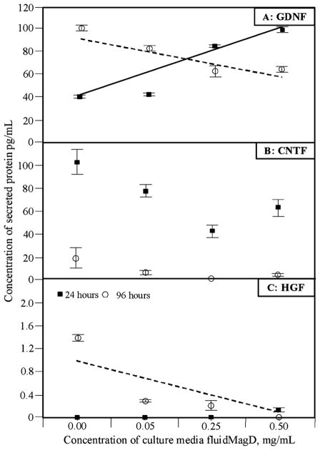
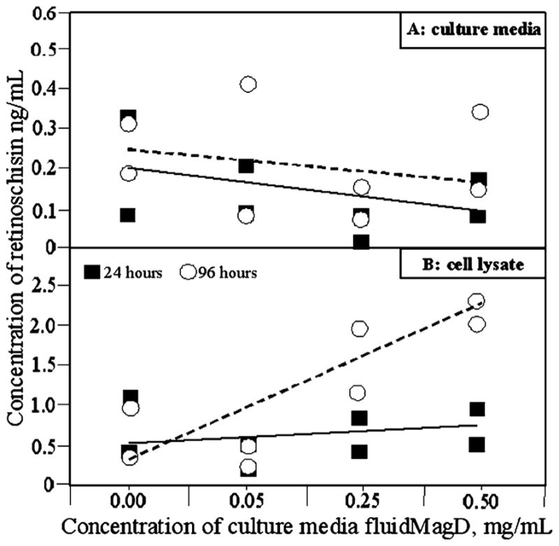
Mesenchymal stem cells (MSCs) have well-established paracrine effects that are proving to be therapeutically useful. This potential is based on the ability of MSCs to secrete a range of neuroprotective and anti-inflammatory molecules. Previous work in our laboratory has demonstrated that intravenous injection of MSCs, treated with superparamagnetic iron oxide nanoparticle fluidMAG-D resulted in enhanced levels of glial-derived neurotrophic factor, ciliary neurotrophic factor, hepatocyte growth factor and interleukin-10 in the dystrophic rat retina. In this present study we investigated whether the concentration of fluidMAG-D in cell culture media affects the secretion of these four molecules . In addition, we assessed the effect of fluidMAG-D concentration on retinoschisin secretion from genetically modified MSCs. ELISA-assayed secretion of these molecules was measured using escalating concentrations of fluidMAG-D which resulted in MSC iron loads of 0, 7, 120, or 274 pg iron oxide per cell respectively. Our results demonstrated glial-derived neurotrophic factor and hepatocyte growth factor secretion was significantly decreased but only at the 96 hour's time-point whereas no statistically significant effect was seen with ciliary neurotrophic factor secretion. Whereas no effect was observed on culture media concentrations of retinoschisin with increasing iron oxide load, a statistically significant increase in cell lysate retinoschisin concentration (p = 0.01) was observed suggesting that increasing fluidMAG-D concentration did increase retinoschisin production but this did not lead to greater secretion. We hypothesize that higher concentrations of iron-oxide nanoparticle fluidMAG-D have an effect on the innate ability of MSCs to secrete therapeutically useful molecules and also on secretion from genetically modified cells. Further work is required to verify these finding using model systems.
间充质干细胞(MSCs)具有已被充分证实的旁分泌作用,事实证明这些作用具有治疗价值。这种潜力基于MSCs分泌一系列神经保护和抗炎分子的能力。我们实验室之前的工作表明,静脉注射经超顺磁性氧化铁纳米颗粒流体MAG-D处理的MSCs,可使营养不良大鼠视网膜中胶质细胞源性神经营养因子、睫状神经营养因子、肝细胞生长因子和白细胞介素-10的水平升高。在本研究中,我们调查了细胞培养基中流体MAG-D的浓度是否会影响这四种分子的分泌。此外,我们评估了流体MAG-D浓度对基因改造的MSCs分泌视网膜蛋白的影响。使用逐步增加的流体MAG-D浓度(分别导致每个细胞的MSC铁负荷为0、7、120或274 pg氧化铁),通过酶联免疫吸附测定法检测这些分子的分泌情况。我们的结果表明,胶质细胞源性神经营养因子和肝细胞生长因子的分泌显著减少,但仅在96小时的时间点出现这种情况,而睫状神经营养因子的分泌未观察到统计学上的显著影响。虽然随着氧化铁负荷增加,未观察到对培养基中视网膜蛋白浓度有影响,但观察到细胞裂解液中视网膜蛋白浓度有统计学上的显著增加(p = 0.01),这表明增加流体MAG-D浓度确实会增加视网膜蛋白的产生,但这并未导致更多的分泌。我们推测,较高浓度的氧化铁纳米颗粒流体MAG-D会对MSCs分泌具有治疗价值分子的固有能力以及基因改造细胞的分泌产生影响。需要进一步开展工作,使用模型系统来验证这些发现。