• 文献检索
  • 文档翻译
  • 深度研究
  • 学术资讯
  • Suppr Zotero 插件Zotero 插件
  • 邀请有礼
  • 套餐&价格
  • 历史记录
应用&插件
Suppr Zotero 插件Zotero 插件浏览器插件Mac 客户端Windows 客户端微信小程序
定价
高级版会员购买积分包购买API积分包
服务
文献检索文档翻译深度研究API 文档MCP 服务
关于我们
关于 Suppr公司介绍联系我们用户协议隐私条款
关注我们

Suppr 超能文献

核心技术专利:CN118964589B侵权必究
粤ICP备2023148730 号-1Suppr @ 2026

文献检索

告别复杂PubMed语法,用中文像聊天一样搜索,搜遍4000万医学文献。AI智能推荐,让科研检索更轻松。

立即免费搜索

文件翻译

保留排版,准确专业,支持PDF/Word/PPT等文件格式,支持 12+语言互译。

免费翻译文档

深度研究

AI帮你快速写综述,25分钟生成高质量综述,智能提取关键信息,辅助科研写作。

立即免费体验

视网膜视蛋白分泌在 Weri-Rb1 细胞中的调控作用:由 F-肌动蛋白和微管细胞骨架。

Regulation of retinoschisin secretion in Weri-Rb1 cells by the F-actin and microtubule cytoskeleton.

机构信息

Jules Stein Eye Institute and Department of Ophthalmology, David Geffen School of Medicine, University of California Los Angeles, Los Angeles, California, United States of America.

出版信息

PLoS One. 2011;6(6):e20707. doi: 10.1371/journal.pone.0020707. Epub 2011 Jun 27.

DOI:10.1371/journal.pone.0020707
PMID:21738583
原文链接:https://pmc.ncbi.nlm.nih.gov/articles/PMC3124475/
Abstract

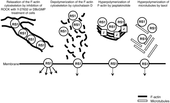
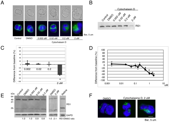
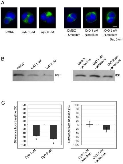
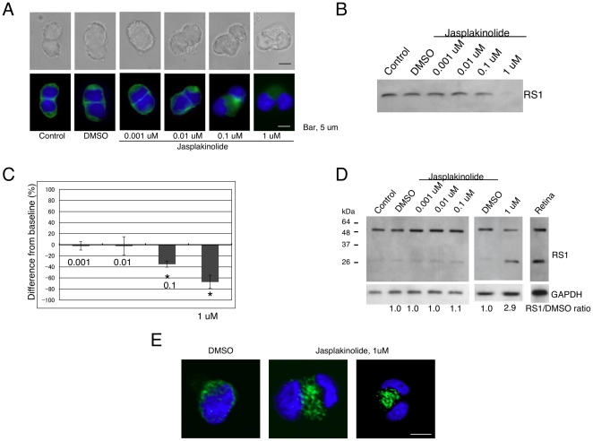
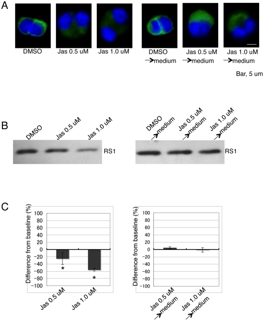
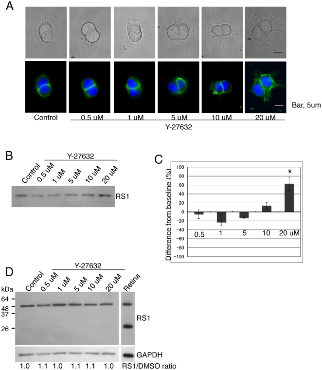
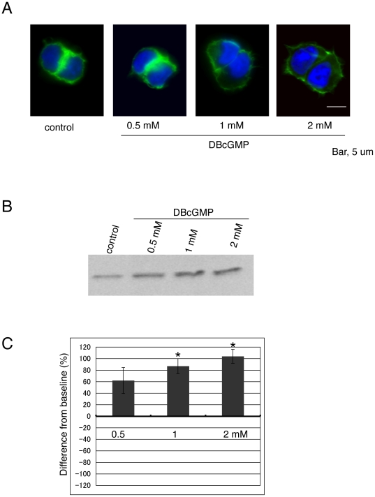
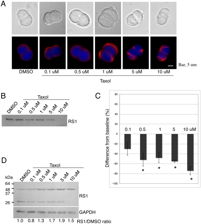
Retinoschisin is encoded by the gene responsible for X-linked retinoschisis (XLRS), an early onset macular degeneration that results in a splitting of the inner layers of the retina and severe loss in vision. Retinoschisin is predominantly expressed and secreted from photoreceptor cells as a homo-oligomer protein; it then associates with the surface of retinal cells and maintains the retina cellular architecture. Many missense mutations in the XLRS1 gene are known to cause intracellular retention of retinoschisin, indicating that the secretion process of the protein is a critical step for its normal function in the retina. However, the molecular mechanisms underlying retinoschisin's secretion remain to be fully elucidated. In this study, we investigated the role of the F-actin cytoskeleton in the secretion of retinoschisin by treating Weri-Rb1 cells, which are known to secrete retinoschisin, with cytochalasin D, jasplakinolide, Y-27632, and dibutyryl cGMP. Our results show that cytochalasin D and jasplakinolide inhibit retinoschisin secretion, whereas Y-27632 and dibutyryl cGMP enhance secretion causing F-actin alterations. We also demonstrate that high concentrations of taxol, which hyperpolymerizes microtubules, inhibit retinoschisin secretion. Our data suggest that retinoschisin secretion is regulated by the F-actin cytoskeleton, that cGMP or inhibition of ROCK alters F-actin structure enhancing the secretion, and that the microtubule cytoskeleton is also involved in this process.

摘要

视锥视蛋白是 X 连锁性视网膜劈裂症(XLRS)相关基因编码的蛋白,XLRS 是一种早期发病的黄斑变性疾病,会导致视网膜内层分裂和严重的视力丧失。视锥视蛋白主要在感光细胞中表达和分泌为同源寡聚体蛋白;然后它与视网膜细胞的表面结合并维持视网膜细胞的结构。已知 XLRS1 基因中的许多错义突变会导致视锥视蛋白在细胞内滞留,表明该蛋白的分泌过程是其在视网膜中正常功能的关键步骤。然而,视锥视蛋白分泌的分子机制仍有待充分阐明。在这项研究中,我们通过用细胞松弛素 D、jasplakinolide、Y-27632 和二丁酰环鸟苷处理已知分泌视锥视蛋白的 Weri-Rb1 细胞,研究了 F-肌动蛋白细胞骨架在视锥视蛋白分泌中的作用。我们的结果表明,细胞松弛素 D 和 jasplakinolide 抑制视锥视蛋白分泌,而 Y-27632 和二丁酰环鸟苷增强分泌导致 F-肌动蛋白结构改变。我们还证明,高浓度的紫杉醇(可使微管高度聚合)抑制视锥视蛋白分泌。我们的数据表明,视锥视蛋白的分泌受 F-肌动蛋白细胞骨架调节,cGMP 或 ROCK 抑制改变 F-肌动蛋白结构,增强分泌,微管细胞骨架也参与该过程。

https://cdn.ncbi.nlm.nih.gov/pmc/blobs/61fc/3124475/e276b350e1ed/pone.0020707.g009.jpg
https://cdn.ncbi.nlm.nih.gov/pmc/blobs/61fc/3124475/1561a004f12e/pone.0020707.g001.jpg
https://cdn.ncbi.nlm.nih.gov/pmc/blobs/61fc/3124475/4c56cc9c54e2/pone.0020707.g002.jpg
https://cdn.ncbi.nlm.nih.gov/pmc/blobs/61fc/3124475/f707a54d0b3e/pone.0020707.g003.jpg
https://cdn.ncbi.nlm.nih.gov/pmc/blobs/61fc/3124475/4a4c08964868/pone.0020707.g004.jpg
https://cdn.ncbi.nlm.nih.gov/pmc/blobs/61fc/3124475/967e6021f7d2/pone.0020707.g005.jpg
https://cdn.ncbi.nlm.nih.gov/pmc/blobs/61fc/3124475/4cd2943bef53/pone.0020707.g006.jpg
https://cdn.ncbi.nlm.nih.gov/pmc/blobs/61fc/3124475/d668f6c12a2c/pone.0020707.g007.jpg
https://cdn.ncbi.nlm.nih.gov/pmc/blobs/61fc/3124475/d4e9b6be44a6/pone.0020707.g008.jpg
https://cdn.ncbi.nlm.nih.gov/pmc/blobs/61fc/3124475/e276b350e1ed/pone.0020707.g009.jpg

相似文献

1
Regulation of retinoschisin secretion in Weri-Rb1 cells by the F-actin and microtubule cytoskeleton.视网膜视蛋白分泌在 Weri-Rb1 细胞中的调控作用:由 F-肌动蛋白和微管细胞骨架。
PLoS One. 2011;6(6):e20707. doi: 10.1371/journal.pone.0020707. Epub 2011 Jun 27.
2
Retinoschisin (RS1), the protein encoded by the X-linked retinoschisis gene, is anchored to the surface of retinal photoreceptor and bipolar cells through its interactions with a Na/K ATPase-SARM1 complex.视网膜分裂蛋白(RS1)是由X连锁视网膜分裂症基因编码的蛋白质,通过与钠钾ATP酶-SARM1复合物相互作用,锚定在视网膜光感受器和双极细胞表面。
J Biol Chem. 2007 Nov 9;282(45):32792-801. doi: 10.1074/jbc.M706321200. Epub 2007 Sep 5.
3
TREK-1 Regulates Cytokine Secretion from Cultured Human Alveolar Epithelial Cells Independently of Cytoskeletal Rearrangements.TREK-1独立于细胞骨架重排调节培养的人肺泡上皮细胞的细胞因子分泌。
PLoS One. 2015 May 22;10(5):e0126781. doi: 10.1371/journal.pone.0126781. eCollection 2015.
4
Retinoschisin, the X-linked retinoschisis protein, is a secreted photoreceptor protein, and is expressed and released by Weri-Rb1 cells.视网膜劈裂蛋白,即X连锁视网膜劈裂症蛋白,是一种分泌型光感受器蛋白,由Weri-Rb1细胞表达并释放。
Hum Mol Genet. 2000 Jul 22;9(12):1873-9. doi: 10.1093/hmg/9.12.1873.
5
Molecular pathology of X linked retinoschisis: mutations interfere with retinoschisin secretion and oligomerisation.X连锁视网膜劈裂症的分子病理学:突变干扰视网膜劈裂蛋白的分泌和寡聚化。
Br J Ophthalmol. 2006 Jan;90(1):81-6. doi: 10.1136/bjo.2005.078048.
6
The X-linked juvenile retinoschisis protein retinoschisin is a novel regulator of mitogen-activated protein kinase signalling and apoptosis in the retina.X连锁青少年视网膜劈裂症蛋白视网膜劈裂蛋白是视网膜中丝裂原活化蛋白激酶信号传导和细胞凋亡的新型调节因子。
J Cell Mol Med. 2017 Apr;21(4):768-780. doi: 10.1111/jcmm.13019. Epub 2016 Dec 20.
7
Defective discoidin domain structure, subunit assembly, and endoplasmic reticulum processing of retinoschisin are primary mechanisms responsible for X-linked retinoschisis.视网膜分裂蛋白的盘状结构域缺陷、亚基组装及内质网加工过程是导致X连锁视网膜劈裂症的主要机制。
J Biol Chem. 2003 Jul 25;278(30):28139-46. doi: 10.1074/jbc.M302464200. Epub 2003 May 13.
8
Pathomechanism of mutated and secreted retinoschisin in X-linked juvenile retinoschisis.X 连锁青年性视网膜劈裂症中突变和分泌的视网膜色素上皮衍生因子的发病机制。
Exp Eye Res. 2018 Dec;177:23-34. doi: 10.1016/j.exer.2018.07.021. Epub 2018 Jul 21.
9
X-linked juvenile retinoschisis: clinical diagnosis, genetic analysis, and molecular mechanisms.X 连锁青少年性视网膜劈裂症:临床诊断、基因分析及分子机制。
Prog Retin Eye Res. 2012 May;31(3):195-212. doi: 10.1016/j.preteyeres.2011.12.002. Epub 2012 Jan 3.
10
The Na/K-ATPase is obligatory for membrane anchorage of retinoschisin, the protein involved in the pathogenesis of X-linked juvenile retinoschisis.钠钾 ATP 酶是视锥视蛋白固定在细胞膜上所必需的,该蛋白与 X 连锁青年性视网膜劈裂症的发病机制有关。
Hum Mol Genet. 2011 Mar 15;20(6):1132-42. doi: 10.1093/hmg/ddq557. Epub 2010 Dec 31.

引用本文的文献

1
Evaluation of mesenchymal stem cells as an model for inherited retinal diseases.间充质干细胞作为遗传性视网膜疾病模型的评估。
Front Cell Dev Biol. 2024 Nov 15;12:1455140. doi: 10.3389/fcell.2024.1455140. eCollection 2024.
2
Effectiveness of Ripasudil, a Rho-Associated Coiled/Coil-Containing Protein Kinase Inhibitor, in Improving Retinoschisis and Cystic-Like Foveal Cavities in Eyes with X-Linked Retinoschisis.Rho相关卷曲螺旋蛋白激酶抑制剂力法司他改善X连锁视网膜劈裂症患者视网膜劈裂和黄斑区囊样腔隙的有效性
Case Rep Ophthalmol. 2020 Aug 6;11(2):411-417. doi: 10.1159/000509261. eCollection 2020 May-Aug.
3
Influence of Iron Oxide Nanoparticles on Innate and Genetically Modified Secretion Profiles of Mesenchymal Stem Cells.

本文引用的文献

1
Cofilin phosphorylation is involved in nitric oxide/cGMP-mediated nociception.丝切蛋白磷酸化参与了一氧化氮/cGMP 介导的痛觉感受。
Biochem Biophys Res Commun. 2009 Dec 25;390(4):1408-13. doi: 10.1016/j.bbrc.2009.10.166. Epub 2009 Nov 5.
2
Mechanisms of biphasic insulin-granule exocytosis - roles of the cytoskeleton, small GTPases and SNARE proteins.双相胰岛素颗粒胞吐作用机制——细胞骨架、小GTP酶和SNARE蛋白的作用
J Cell Sci. 2009 Apr 1;122(Pt 7):893-903. doi: 10.1242/jcs.034355.
3
Role of the Rho-ROCK (Rho-associated kinase) signaling pathway in the regulation of pancreatic beta-cell function.
氧化铁纳米颗粒对间充质干细胞固有及基因改造分泌谱的影响
IEEE Trans Magn. 2013 Jan 1;49(1):389-393. doi: 10.1109/TMAG.2012.2225829.
Rho-ROCK(Rho相关激酶)信号通路在胰腺β细胞功能调节中的作用。
Endocrinology. 2009 May;150(5):2072-9. doi: 10.1210/en.2008-1135. Epub 2008 Dec 23.
4
Retinoschisin, a new binding partner for L-type voltage-gated calcium channels in the retina.视网膜分裂蛋白,视网膜中L型电压门控钙通道的一种新结合伴侣。
J Biol Chem. 2009 Feb 6;284(6):3966-75. doi: 10.1074/jbc.M806333200. Epub 2008 Dec 11.
5
Clustering of endocytic organelles in parental and drug-resistant myeloid leukaemia cell lines lacking centrosomally organised microtubule arrays.在缺乏中心体组织微管阵列的亲代和耐药性髓系白血病细胞系中,内吞细胞器的聚集。
Int J Biochem Cell Biol. 2008;40(10):2240-52. doi: 10.1016/j.biocel.2008.03.004. Epub 2008 Mar 16.
6
Circadian regulation of retinoschisin in the chick retina.雏鸡视网膜中视网膜分裂蛋白的昼夜节律调节
Invest Ophthalmol Vis Sci. 2008 Apr;49(4):1615-21. doi: 10.1167/iovs.07-1189.
7
Ca2+ -dependent regulation of phototransduction.光转导的钙离子依赖性调节
Photochem Photobiol. 2008 Jul-Aug;84(4):903-10. doi: 10.1111/j.1751-1097.2008.00323.x. Epub 2008 Mar 12.
8
Proteomics of photoreceptor outer segments identifies a subset of SNARE and Rab proteins implicated in membrane vesicle trafficking and fusion.光感受器外段的蛋白质组学鉴定出了一组与膜泡运输和融合相关的SNARE蛋白和Rab蛋白。
Mol Cell Proteomics. 2008 Jun;7(6):1053-66. doi: 10.1074/mcp.M700571-MCP200. Epub 2008 Jan 31.
9
NO/cGMP-dependent modulation of synaptic transmission.一氧化氮/环磷酸鸟苷依赖性的突触传递调节
Handb Exp Pharmacol. 2008(184):529-60. doi: 10.1007/978-3-540-74805-2_16.
10
Cytoskeletal control of vesicle transport and exocytosis in chromaffin cells.嗜铬细胞中囊泡运输和胞吐作用的细胞骨架控制
Acta Physiol (Oxf). 2008 Feb;192(2):165-72. doi: 10.1111/j.1748-1716.2007.01808.x. Epub 2007 Nov 16.