Vandergugten Simon, Traore Sidi Yaya, Cartiaux Olivier, Lecouvet Frédéric, Galant Christine, Docquier Pierre-Louis
Computer Assisted and Robotic Surgery (CARS), Institut de Recherche Expérimentale et Clinique, Université Catholique de Louvain, avenue Mounier 53, 1200 Brussels, Belgium ; Service de Chirurgie Orthopédique et Traumatologique, Cliniques Universitaires Saint-Luc, avenue Hippocrate 10, 1200 Brussels, Belgium.
Computer Assisted and Robotic Surgery (CARS), Institut de Recherche Expérimentale et Clinique, Université Catholique de Louvain, avenue Mounier 53, 1200 Brussels, Belgium.
Sarcoma. 2014;2014:967848. doi: 10.1155/2014/967848. Epub 2014 May 26.
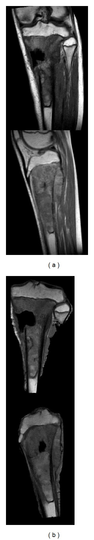
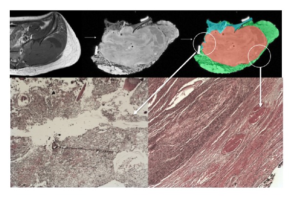
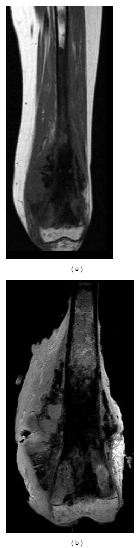
In 12 patients operated on for bone sarcoma resection, a postoperative magnetic resonance imaging of the resection specimens was obtained in order to assess the surgical margins. Margins were classified according to MRI in R0, R1, and R2 by three independent observers: a radiologist and two orthopaedic surgeons. Final margin evaluation (R0, R1, and R2) was assessed by a confirmed pathologist. Agreement for margin evaluation between the pathologist and the radiologist was perfect (κ = 1). Agreement between the pathologist and an experienced orthopaedic surgeon was very good while it was fair between the pathologist and a junior orthopaedic surgeon. MRI should be considered as a tool to give quick information about the adequacy of margins and to help the pathologist to focus on doubtful areas and to spare time in specimen analysis. But it may not replace the pathological evaluation that gives additional information about tumor necrosis. This study shows that MRI extemporaneous analysis of a resection specimen may be efficient in bone tumor oncologic surgery, if made by an experienced radiologist with perfect agreement with the pathologist.
在12例接受骨肉瘤切除手术的患者中,术后对切除标本进行了磁共振成像,以评估手术切缘。由一名放射科医生和两名骨科医生这三位独立观察者根据磁共振成像将切缘分为R0、R1和R2三类。最终的切缘评估(R0、R1和R2)由一名确诊病理学家进行。病理学家与放射科医生之间在切缘评估方面的一致性极佳(κ = 1)。病理学家与一名经验丰富的骨科医生之间的一致性很好,而与一名初级骨科医生之间的一致性一般。应将磁共振成像视为一种工具,用于快速提供有关切缘充分性的信息,并帮助病理学家关注可疑区域,节省标本分析时间。但它可能无法取代病理评估,病理评估能提供有关肿瘤坏死的额外信息。本研究表明,如果由经验丰富的放射科医生进行,且与病理学家的一致性良好,那么对切除标本进行磁共振成像即时分析在骨肿瘤肿瘤外科手术中可能是有效的。