• 文献检索
  • 文档翻译
  • 深度研究
  • 学术资讯
  • Suppr Zotero 插件Zotero 插件
  • 邀请有礼
  • 套餐&价格
  • 历史记录
应用&插件
Suppr Zotero 插件Zotero 插件浏览器插件Mac 客户端Windows 客户端微信小程序
定价
高级版会员购买积分包购买API积分包
服务
文献检索文档翻译深度研究API 文档MCP 服务
关于我们
关于 Suppr公司介绍联系我们用户协议隐私条款
关注我们

Suppr 超能文献

核心技术专利:CN118964589B侵权必究
粤ICP备2023148730 号-1Suppr @ 2026

文献检索

告别复杂PubMed语法,用中文像聊天一样搜索,搜遍4000万医学文献。AI智能推荐,让科研检索更轻松。

立即免费搜索

文件翻译

保留排版,准确专业,支持PDF/Word/PPT等文件格式,支持 12+语言互译。

免费翻译文档

深度研究

AI帮你快速写综述,25分钟生成高质量综述,智能提取关键信息,辅助科研写作。

立即免费体验

利用离体磁共振成像评估舌鳞状细胞癌的手术切缘

Evaluation of tongue squamous cell carcinoma resection margins using ex-vivo MR.

作者信息

Steens Stefan C A, Bekers Elise M, Weijs Willem L J, Litjens Geert J S, Veltien Andor, Maat Arie, van den Broek Guido B, van der Laak Jeroen A W M, Fütterer Jürgen J, van der Kaa Christina A Hulsbergen, Merkx Matthias A W, Takes Robert P

机构信息

Department of Radiology and Nuclear Medicine, Radboud University Medical Center, P.O. Box 9101, 6500 HB, Nijmegen, The Netherlands.

Department of Pathology, Radboud University Medical Center, Nijmegen, The Netherlands.

出版信息

Int J Comput Assist Radiol Surg. 2017 May;12(5):821-828. doi: 10.1007/s11548-017-1524-6. Epub 2017 Jan 27.

DOI:10.1007/s11548-017-1524-6
PMID:28130702
原文链接:https://pmc.ncbi.nlm.nih.gov/articles/PMC5420007/
Abstract

PURPOSE

Purpose of this feasibility study was (1) to evaluate whether application of ex-vivo 7T MR of the resected tongue specimen containing squamous cell carcinoma may provide information on the resection margin status and (2) to evaluate the research and developmental issues that have to be solved for this technique to have the beneficial impact on clinical outcome that we expect: better oncologic and functional outcomes, better quality of life, and lower costs.

METHODS

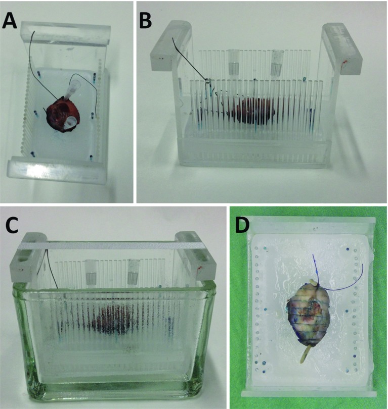
We performed a non-blinded validation of ex-vivo 7T MR to detect the tongue squamous cell carcinoma and resection margin in 10 fresh tongue specimens using histopathology as gold standard.

RESULTS

In six of seven specimens with a histopathologically determined invasion depth of the tumor of [Formula: see text] mm, the tumor could be recognized on MR, with a resection margin within a 2 mm range as compared to histopathology. In three specimens with an invasion depth of [Formula: see text] mm, the tumor was not visible on MR. Technical limitations mainly included scan time, image resolution, and the fact that we used a less available small-bore 7T MR machine.

CONCLUSION

Ex-vivo 7T probably will have a low negative predictive value but a high positive predictive value, meaning that in tumors thicker than a few millimeters we expect to be able to predict whether the resection margin is too small. A randomized controlled trial needs to be performed to show our hypothesis: better oncologic and functional outcomes, better quality of life, and lower costs.

摘要

目的

本可行性研究的目的是(1)评估对含有鳞状细胞癌的切除舌标本进行离体7T磁共振成像(MR)是否能提供切缘状态的信息,以及(2)评估为使该技术对临床结局产生预期的有益影响(更好的肿瘤学和功能结局、更高的生活质量以及更低的成本)而必须解决的研发问题。

方法

我们对10个新鲜舌标本进行了离体7T MR检测舌鳞状细胞癌和切缘的非盲法验证,以组织病理学作为金标准。

结果

在7个组织病理学确定肿瘤浸润深度为[公式:见正文]毫米的标本中,有6个标本在MR上可识别出肿瘤,与组织病理学相比,切缘在2毫米范围内。在3个浸润深度为[公式:见正文]毫米的标本中,肿瘤在MR上不可见。技术限制主要包括扫描时间、图像分辨率,以及我们使用的是一台可用性较低的小口径7T MR机器这一事实。

结论

离体7T MR可能具有较低的阴性预测值但较高的阳性预测值,这意味着在厚度超过几毫米的肿瘤中,我们预计能够预测切缘是否过小。需要进行一项随机对照试验来验证我们的假设:更好的肿瘤学和功能结局、更高的生活质量以及更低的成本。

https://cdn.ncbi.nlm.nih.gov/pmc/blobs/a756/5420007/feb3b676278f/11548_2017_1524_Fig3_HTML.jpg
https://cdn.ncbi.nlm.nih.gov/pmc/blobs/a756/5420007/f57c8db2e96f/11548_2017_1524_Fig1_HTML.jpg
https://cdn.ncbi.nlm.nih.gov/pmc/blobs/a756/5420007/a54ef29715cc/11548_2017_1524_Fig2_HTML.jpg
https://cdn.ncbi.nlm.nih.gov/pmc/blobs/a756/5420007/feb3b676278f/11548_2017_1524_Fig3_HTML.jpg
https://cdn.ncbi.nlm.nih.gov/pmc/blobs/a756/5420007/f57c8db2e96f/11548_2017_1524_Fig1_HTML.jpg
https://cdn.ncbi.nlm.nih.gov/pmc/blobs/a756/5420007/a54ef29715cc/11548_2017_1524_Fig2_HTML.jpg
https://cdn.ncbi.nlm.nih.gov/pmc/blobs/a756/5420007/feb3b676278f/11548_2017_1524_Fig3_HTML.jpg

相似文献

1
Evaluation of tongue squamous cell carcinoma resection margins using ex-vivo MR.利用离体磁共振成像评估舌鳞状细胞癌的手术切缘
Int J Comput Assist Radiol Surg. 2017 May;12(5):821-828. doi: 10.1007/s11548-017-1524-6. Epub 2017 Jan 27.
2
Feasibility of a Novel 3D Ultrasound Imaging Technique for Intraoperative Margin Assessment during Tongue Cancer Surgery.一种新型三维超声成像技术在舌癌手术中用于术中切缘评估的可行性研究。
Curr Oncol. 2024 Aug 1;31(8):4414-4431. doi: 10.3390/curroncol31080330.
3
Optical coherence tomography in the assessment of oral squamous cell carcinoma resection margins.光学相干断层扫描在口腔鳞状细胞癌切除边缘评估中的应用
Photodiagnosis Photodyn Ther. 2016 Mar;13:211-217. doi: 10.1016/j.pdpdt.2015.07.170. Epub 2015 Jul 22.
4
Intra-operative intaoral ultrasonograpy ensures free deep resection margins in carcinoma tongue: A pilot study.术中口腔内超声检查确保舌癌的无瘤深切缘:一项初步研究。
J Cancer Res Ther. 2023 Jan 1;19(Suppl 2):S877-S880. doi: 10.4103/jcrt.jcrt_2496_22. Epub 2023 May 3.
5
Toward assessment of resection margins using hyperspectral diffuse reflection imaging (400-1,700 nm) during tongue cancer surgery.利用舌癌手术期间的高光谱漫反射成像(400-1700nm)评估切缘。
Lasers Surg Med. 2020 Jul;52(6):496-502. doi: 10.1002/lsm.23161. Epub 2019 Sep 15.
6
Correlation between clinical and MRI assessment of depth of invasion in oral tongue squamous cell carcinoma.口腔舌鳞状细胞癌侵袭深度的临床评估与MRI评估之间的相关性
J Otolaryngol Head Neck Surg. 2016 Nov 22;45(1):61. doi: 10.1186/s40463-016-0172-0.
7
Feasibility study of ultrasound-guided resection of tongue cancer with immediate specimen examination to improve margin control - Comparison with conventional treatment.超声引导下舌癌切除术并即时标本检查以改善切缘控制的可行性研究——与传统治疗的比较
Oral Oncol. 2021 May;116:105249. doi: 10.1016/j.oraloncology.2021.105249. Epub 2021 Mar 26.
8
The role of magnetic resonance imaging in assessing the extent of tongue squamous cell carcinoma: A prospective cohort study.磁共振成像在评估舌鳞癌范围中的作用:一项前瞻性队列研究。
J Stomatol Oral Maxillofac Surg. 2022 Nov;123(6):e822-e827. doi: 10.1016/j.jormas.2022.03.006. Epub 2022 Mar 4.
9
Application and accuracy of ultrasound-guided resections of tongue cancer.超声引导下舌癌切除术的应用及准确性。
Oral Oncol. 2022 Oct;133:106023. doi: 10.1016/j.oraloncology.2022.106023. Epub 2022 Jul 25.
10
Assessment of surgical tumor-free resection margins in fresh squamous-cell carcinoma resection specimens of the tongue using a clinical MRI system.使用临床MRI系统评估舌部新鲜鳞状细胞癌切除标本的手术无瘤切缘。
Head Neck. 2020 Aug;42(8):2039-2049. doi: 10.1002/hed.26125. Epub 2020 Mar 2.

引用本文的文献

1
Tongue Squamous Cell Carcinoma Prognosis Can Be Effectively Predicted by LncRNA LIPH4: A Prospective Study.长链非编码RNA LIPH4可有效预测舌鳞状细胞癌预后:一项前瞻性研究
Int J Gen Med. 2024 Sep 16;17:4119-4126. doi: 10.2147/IJGM.S474142. eCollection 2024.
2
Feasibility of a Novel 3D Ultrasound Imaging Technique for Intraoperative Margin Assessment during Tongue Cancer Surgery.一种新型三维超声成像技术在舌癌手术中用于术中切缘评估的可行性研究。
Curr Oncol. 2024 Aug 1;31(8):4414-4431. doi: 10.3390/curroncol31080330.
3
Feasibility of an MR-based digital specimen for tongue cancer resection specimens: a novel approach for margin evaluation.

本文引用的文献

1
Resection margins in oral cancer surgery: Room for improvement.口腔癌手术中的切缘:仍有改进空间。
Head Neck. 2016 Apr;38 Suppl 1:E2197-203. doi: 10.1002/hed.24075. Epub 2015 Jun 15.
2
Diagnostic Accuracy of Preoperative Gadoxetic Acid-enhanced 3-T MR Imaging for Malignant Liver Lesions by Using Ex Vivo MR Imaging-matched Pathologic Findings as the Reference Standard.以离体磁共振成像匹配的病理结果为参考标准评估术前钆塞酸二钠增强3T磁共振成像对肝脏恶性病变的诊断准确性
Radiology. 2015 Sep;276(3):775-86. doi: 10.1148/radiol.2015142069. Epub 2015 Apr 15.
3
Ultra-high-resolution MR imaging of esophageal carcinoma at ultra-high field strength (7.0T) ex vivo: correlation with histopathologic findings.
基于磁共振成像的舌癌切除标本数字标本的可行性:一种切缘评估的新方法。
Front Oncol. 2024 Mar 28;14:1342857. doi: 10.3389/fonc.2024.1342857. eCollection 2024.
4
3D Ultrasound and MRI in Assessing Resection Margins during Tongue Cancer Surgery: A Research Protocol for a Clinical Diagnostic Accuracy Study.3D超声和MRI在舌癌手术中评估切缘的应用:一项临床诊断准确性研究的研究方案
J Imaging. 2023 Aug 28;9(9):174. doi: 10.3390/jimaging9090174.
5
Image-Guided Intraoperative Assessment of Surgical Margins in Oral Cavity Squamous Cell Cancer: A Diagnostic Test Accuracy Review.口腔鳞状细胞癌手术切缘的图像引导术中评估:诊断试验准确性综述
Diagnostics (Basel). 2023 May 25;13(11):1846. doi: 10.3390/diagnostics13111846.
6
Intraoperative Imaging Techniques to Improve Surgical Resection Margins of Oropharyngeal Squamous Cell Cancer: A Comprehensive Review of Current Literature.改善口咽鳞状细胞癌手术切缘的术中成像技术:当前文献综述
Cancers (Basel). 2023 Jan 31;15(3):896. doi: 10.3390/cancers15030896.
7
3D Ultrasound versus Computed Tomography for Tumor Volume Measurement Compared to Gross Pathology-A Pilot Study on an Animal Model.与大体病理学相比,3D超声与计算机断层扫描在肿瘤体积测量中的应用——一项动物模型的初步研究
J Imaging. 2022 Dec 19;8(12):329. doi: 10.3390/jimaging8120329.
8
Frozen Section Analysis and Real-Time Magnetic Resonance Imaging of Surgical Specimen Oriented on 3D Printed Tongue Model to Assess Surgical Margins in Oral Tongue Carcinoma: Preliminary Results.基于3D打印舌模型的手术标本冷冻切片分析及实时磁共振成像评估口腔舌癌手术切缘:初步结果
Front Oncol. 2021 Dec 9;11:735002. doi: 10.3389/fonc.2021.735002. eCollection 2021.
9
RF Coil Setup for P MRSI in Tongue Cancer at 7 T.7T下舌癌磷磁共振波谱成像的射频线圈设置
Front Neurol. 2021 Nov 2;12:695202. doi: 10.3389/fneur.2021.695202. eCollection 2021.
10
Intraoperative imaging in pathology-assisted surgery.术中病理辅助手术的影像学检查
Nat Biomed Eng. 2022 May;6(5):503-514. doi: 10.1038/s41551-021-00808-8. Epub 2021 Nov 8.
超高场强(7.0T)离体食管癌的超高分辨率磁共振成像:与组织病理学结果的相关性
Magn Reson Imaging. 2015 May;33(4):413-9. doi: 10.1016/j.mri.2014.11.005. Epub 2015 Jan 30.
4
Gastric Carcinoma: Ex Vivo MR Imaging at 7.0 T-Correlation with Histopathologic Findings.胃癌:7.0T 外生 MR 成像与组织病理学发现的相关性。
Radiology. 2015 Jun;275(3):841-8. doi: 10.1148/radiol.14141878. Epub 2015 Jan 14.
5
Analysis of glioblastoma tumor coverage by oncolytic virus-loaded neural stem cells using MRI-based tracking and histological reconstruction.使用基于磁共振成像的追踪和组织学重建分析溶瘤病毒负载的神经干细胞对胶质母细胞瘤的肿瘤覆盖情况。
Cancer Gene Ther. 2015 Jan;22(1):55-61. doi: 10.1038/cgt.2014.72. Epub 2014 Dec 19.
6
MR elastography and diffusion-weighted imaging of ex vivo prostate cancer: quantitative comparison to histopathology.离体前列腺癌的磁共振弹性成像与扩散加权成像:与组织病理学的定量比较
NMR Biomed. 2015 Jan;28(1):89-100. doi: 10.1002/nbm.3203. Epub 2014 Nov 10.
7
Assessment of change in prostate volume and shape following surgical resection through co-registration of in-vivo MRI and fresh specimen ex-vivo MRI.通过体内 MRI 和新鲜标本离体 MRI 的配准评估手术切除后前列腺体积和形状的变化。
Clin Radiol. 2014 Oct;69(10):e398-403. doi: 10.1016/j.crad.2014.06.012. Epub 2014 Jul 22.
8
MRI evaluation of resection margins in bone tumour surgery.骨肿瘤手术切缘的磁共振成像评估
Sarcoma. 2014;2014:967848. doi: 10.1155/2014/967848. Epub 2014 May 26.
9
Esophageal carcinoma: Evaluation with q-space diffusion-weighted MR imaging ex vivo.食管癌:离体q空间扩散加权磁共振成像评估
Magn Reson Med. 2015 Jun;73(6):2262-73. doi: 10.1002/mrm.25334. Epub 2014 Jun 19.
10
Registration of in vivo prostate MRI and pseudo-whole mount histology using Local Affine Transformations guided by Internal Structures (LATIS).使用由内部结构引导的局部仿射变换(LATIS)对体内前列腺MRI和伪全层组织学进行配准。
J Magn Reson Imaging. 2015 Apr;41(4):1104-14. doi: 10.1002/jmri.24629. Epub 2014 Apr 4.