Murai Yasuo, Mizunari Takayuki, Koketsu Kenta, Tateyama Kojiro, Kobayashi Shiro, Morita Akio, Teramoto Akira
Department of Neurosurgery, Nippon Medical School.
Neurol Med Chir (Tokyo). 2015;55(8):683-8. doi: 10.2176/nmc.tn.2013-0249. Epub 2014 Jul 4.
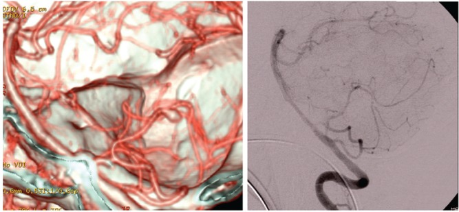
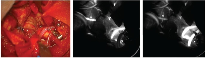
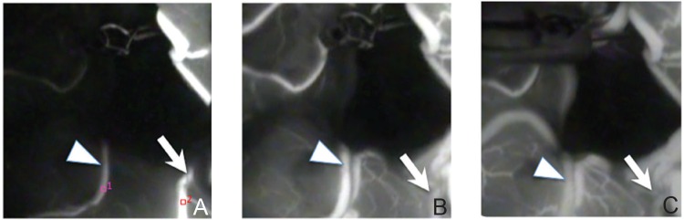
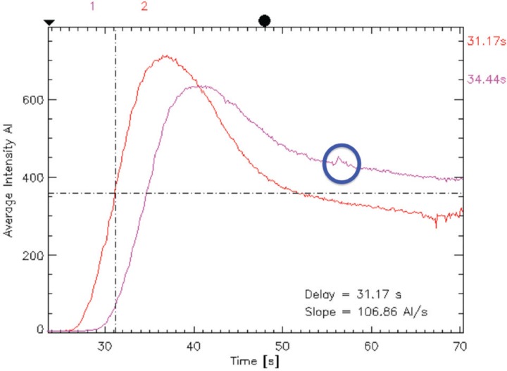
Confirming the patency of the proximal parent and distal artery is necessary in cerebral aneurysm surgery. To understand the relationship between the parent and distal arteries of the aneurysm, the blood vessels running through the subarachnoid space should be extensively dissected, which is time consuming. To examine the efficacy of a temporary clip with indocyanine green (ICG) technique, in which the parent artery is temporarily occluded using a temporary clip, an ICG videoangiography (ICGVAG) is performed to clarify the relationship between the distal artery and the proximal parent artery. Three patients with a distal aneurysm. This technique was used to confirm the connection of the parent and the distal artery in distal aneurysms. With regard to middle cerebral artery (MCA), the procedure is conducted as follows. First, the M2 within the Sylvian fissure is investigated to ensure the absence of atherosclerosis and perforators and that this vessel could undergo occlusion by temporary clipping. The subarachnoid space surrounding the distal artery of the lesion site suspected of an existent aneurysm is dissected. The image range of the ICGVAG is set sufficiently wide to accommodate the possibility that the distal artery is not the artery that was anticipated. Subsequently, after the temporary clip occlusion is completed, the ICGVAG is recorded. In the three distal aneurysms, the relationship between the aneurysm, the distal artery, and the parent artery was confirmed. This method was useful, suggesting that unnecessary dissection in the subarachnoid space might be reduced.
在脑动脉瘤手术中,确认近端供血动脉和远端动脉的通畅性是必要的。为了解动脉瘤的供血动脉与远端动脉之间的关系,需要广泛解剖穿过蛛网膜下腔的血管,这很耗时。为了检验使用吲哚菁绿(ICG)技术的临时夹闭的效果,即使用临时夹暂时夹闭供血动脉,需进行ICG视频血管造影(ICGVAG)以明确远端动脉与近端供血动脉之间的关系。三名患有远端动脉瘤的患者。该技术用于确认远端动脉瘤中供血动脉与远端动脉的连接情况。对于大脑中动脉(MCA),操作如下。首先,探查外侧裂内的M2段,以确保无动脉粥样硬化和穿支血管,并且该血管可通过临时夹闭进行阻断。解剖疑似存在动脉瘤的病变部位远端动脉周围的蛛网膜下腔。将ICGVAG的图像范围设置得足够宽,以应对远端动脉并非预期动脉的可能性。随后,在完成临时夹闭后,记录ICGVAG。在这三个远端动脉瘤中,确认了动脉瘤、远端动脉和供血动脉之间的关系。该方法很有用,表明可能减少在蛛网膜下腔进行不必要的解剖。