• 文献检索
  • 文档翻译
  • 深度研究
  • 学术资讯
  • Suppr Zotero 插件Zotero 插件
  • 邀请有礼
  • 套餐&价格
  • 历史记录
应用&插件
Suppr Zotero 插件Zotero 插件浏览器插件Mac 客户端Windows 客户端微信小程序
定价
高级版会员购买积分包购买API积分包
服务
文献检索文档翻译深度研究API 文档MCP 服务
关于我们
关于 Suppr公司介绍联系我们用户协议隐私条款
关注我们

Suppr 超能文献

核心技术专利:CN118964589B侵权必究
粤ICP备2023148730 号-1Suppr @ 2026

文献检索

告别复杂PubMed语法,用中文像聊天一样搜索,搜遍4000万医学文献。AI智能推荐,让科研检索更轻松。

立即免费搜索

文件翻译

保留排版,准确专业,支持PDF/Word/PPT等文件格式,支持 12+语言互译。

免费翻译文档

深度研究

AI帮你快速写综述,25分钟生成高质量综述,智能提取关键信息,辅助科研写作。

立即免费体验

视网膜血管几何形状变化与糖尿病视网膜病变的关系及其在进展预测中的作用——一项探索性研究。

The association between retinal vascular geometry changes and diabetic retinopathy and their role in prediction of progression--an exploratory study.

机构信息

Sunderland Eye Infirmary - Queen Alexandra Road, Sunderland SR2 9HP, UK.

出版信息

BMC Ophthalmol. 2014 Jul 7;14:89. doi: 10.1186/1471-2415-14-89.

DOI:10.1186/1471-2415-14-89
PMID:25001248
原文链接:https://pmc.ncbi.nlm.nih.gov/articles/PMC4094636/
Abstract

BACKGROUND

The study describes the relationship of retinal vascular geometry (RVG) to severity of diabetic retinopathy (DR), and its predictive role for subsequent development of proliferative diabetic retinopathy (PDR).

METHODS

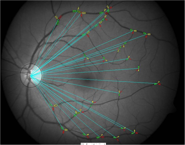
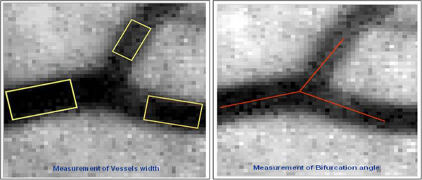
The research project comprises of two stages. Firstly, a comparative study of diabetic patients with different grades of DR. (No DR: Minimal non-proliferative DR: Severe non-proliferative DR: PDR) (10:10: 12: 19). Analysed RVG features including vascular widths and branching angles were compared between patient cohorts. A preliminary statistical model for determination of the retinopathy grade of patients, using these features, is presented. Secondly, in a longitudinal predictive study, RVG features were analysed for diabetic patients with progressive DR over 7 years. RVG at baseline was examined to determine risk for subsequent PDR development.

RESULTS

In the comparative study, increased DR severity was associated with gradual vascular dilatation (p = 0.000), and widening of the bifurcating angle (p = 0.000) with increase in smaller-child-vessel branching angle (p = 0.027). Type 2 diabetes and increased diabetes duration were associated with increased vascular width (p = <0.05 In the predictive study, at baseline, reduced small-child vascular width (OR = 0.73 (95% CI 0.58-0.92)), was predictive of future progression to PDR.

CONCLUSIONS

The study findings suggest that RVG alterations can act as novel markers indicative of progression of DR severity and establishment of PDR. RVG may also have a potential predictive role in determining the risk of future retinopathy progression.

摘要

背景

本研究描述了视网膜血管几何形状(RVG)与糖尿病视网膜病变(DR)严重程度的关系,以及其对增生性糖尿病视网膜病变(PDR)后续发展的预测作用。

方法

该研究项目包括两个阶段。首先,对不同程度 DR 的糖尿病患者进行了比较研究。(无 DR:轻度非增生性 DR:重度非增生性 DR:PDR)(10:10:12:19)。比较了患者队列之间的血管宽度和分支角度等 RVG 特征。提出了一个使用这些特征来确定患者视网膜病变程度的初步统计模型。其次,在一项纵向预测研究中,对 DR 进展的糖尿病患者进行了 RVG 特征分析。检查基线时的 RVG,以确定随后发生 PDR 的风险。

结果

在比较研究中,随着 DR 严重程度的增加,血管逐渐扩张(p = 0.000),分叉角度变宽(p = 0.000),与较小儿童血管分支角度增加(p = 0.027)有关。2 型糖尿病和糖尿病病程延长与血管宽度增加有关(p <0.05)。在预测研究中,基线时,小儿童血管宽度减小(OR = 0.73(95%CI 0.58-0.92)),预示着未来进展为 PDR。

结论

研究结果表明,RVG 改变可作为提示 DR 严重程度进展和 PDR 建立的新型标志物。RVG 也可能在确定未来视网膜病变进展的风险方面具有潜在的预测作用。

https://cdn.ncbi.nlm.nih.gov/pmc/blobs/2d3a/4094636/9b743bbc26d4/1471-2415-14-89-2.jpg
https://cdn.ncbi.nlm.nih.gov/pmc/blobs/2d3a/4094636/ab638e6596a8/1471-2415-14-89-1.jpg
https://cdn.ncbi.nlm.nih.gov/pmc/blobs/2d3a/4094636/9b743bbc26d4/1471-2415-14-89-2.jpg
https://cdn.ncbi.nlm.nih.gov/pmc/blobs/2d3a/4094636/ab638e6596a8/1471-2415-14-89-1.jpg
https://cdn.ncbi.nlm.nih.gov/pmc/blobs/2d3a/4094636/9b743bbc26d4/1471-2415-14-89-2.jpg

相似文献

1
The association between retinal vascular geometry changes and diabetic retinopathy and their role in prediction of progression--an exploratory study.视网膜血管几何形状变化与糖尿病视网膜病变的关系及其在进展预测中的作用——一项探索性研究。
BMC Ophthalmol. 2014 Jul 7;14:89. doi: 10.1186/1471-2415-14-89.
2
Peripheral Lesions Identified on Ultrawide Field Imaging Predict Increased Risk of Diabetic Retinopathy Progression over 4 Years.超广角成像检查发现的周边病变预示着糖尿病视网膜病变在 4 年内进展的风险增加。
Ophthalmology. 2015 May;122(5):949-56. doi: 10.1016/j.ophtha.2015.01.008. Epub 2015 Feb 19.
3
The Relationship of Retinal Vessel Geometric Characteristics to the Incidence and Progression of Diabetic Retinopathy.视网膜血管几何特征与糖尿病视网膜病变的发生和进展的关系。
Ophthalmology. 2018 Nov;125(11):1784-1792. doi: 10.1016/j.ophtha.2018.04.023. Epub 2018 May 18.
4
Retinal main vessel calibers and systemic markers for long-term development of proliferative diabetic retinopathy.视网膜主血管口径和全身标志物与增生性糖尿病视网膜病变的长期发展。
Acta Ophthalmol. 2024 Jun;102(4):448-454. doi: 10.1111/aos.15780. Epub 2023 Oct 7.
5
Early risk stratification in pediatric type 1 diabetes.儿童1型糖尿病的早期风险分层
Acta Ophthalmol. 2015 Mar;93 Thesis 1:1-19. doi: 10.1111/aos.12702.
6
Quantitative Assessment of the Severity of Diabetic Retinopathy.糖尿病视网膜病变严重程度的定量评估。
Am J Ophthalmol. 2020 Oct;218:342-352. doi: 10.1016/j.ajo.2020.05.021. Epub 2020 May 22.
7
Linking Retinal Microvasculature Features With Severity of Diabetic Retinopathy Using Optical Coherence Tomography Angiography.使用光学相干断层扫描血管造影术将视网膜微血管特征与糖尿病性视网膜病变的严重程度相联系。
Invest Ophthalmol Vis Sci. 2016 Jul 1;57(9):OCT519-25. doi: 10.1167/iovs.15-18901.
8
Progressive Retinal Vasodilation in Patients With Type 1 Diabetes: A Longitudinal Study of Retinal Vascular Geometry.1型糖尿病患者视网膜血管的渐进性扩张:视网膜血管几何形态的纵向研究
Invest Ophthalmol Vis Sci. 2017 May 1;58(5):2503-2509. doi: 10.1167/iovs.16-21015.
9
Retinal vascular geometry and its association to microvascular complications in patients with type 1 diabetes: the Danish Cohort of Pediatric Diabetes 1987 (DCPD1987).1型糖尿病患者的视网膜血管几何形态及其与微血管并发症的关联:1987年丹麦儿童糖尿病队列研究(DCPD1987)
Graefes Arch Clin Exp Ophthalmol. 2017 Feb;255(2):293-299. doi: 10.1007/s00417-016-3454-3. Epub 2016 Aug 12.
10
Retinal vascular geometry and 6 year incidence and progression of diabetic retinopathy.视网膜血管几何形态与糖尿病视网膜病变 6 年的发生率和进展。
Diabetologia. 2017 Sep;60(9):1770-1781. doi: 10.1007/s00125-017-4333-0. Epub 2017 Jun 16.

引用本文的文献

1
Robust semi-automatic vessel tracing in the human retinal image by an instance segmentation neural network.通过实例分割神经网络在人类视网膜图像中进行稳健的半自动血管追踪
Sci Adv. 2025 Apr 4;11(14):eado8268. doi: 10.1126/sciadv.ado8268.
2
Peripheral retinal vessel whitening in patients with diabetes mellitus.糖尿病患者的周边视网膜血管变白。
Sci Rep. 2023 May 17;13(1):7981. doi: 10.1038/s41598-023-35124-5.
3
Optic disc and retinal vascular features in first 6 years of Chinese children.中国儿童前6年的视盘和视网膜血管特征

本文引用的文献

1
Early neural and vascular changes in the adolescent type 1 and type 2 diabetic retina.青少年 1 型和 2 型糖尿病视网膜的早期神经和血管变化。
Retina. 2012 Jan;32(1):92-102. doi: 10.1097/IAE.0b013e318219deac.
2
Retinal vascular geometry predicts incident retinopathy in young people with type 1 diabetes: a prospective cohort study from adolescence.视网膜血管几何形态可预测青少年 1 型糖尿病患者的视网膜病变发生:一项前瞻性队列研究。
Diabetes Care. 2011 Jul;34(7):1622-7. doi: 10.2337/dc10-2419. Epub 2011 May 18.
3
Retinal venular diameter as an early indicator of progression to proliferative diabetic retinopathy with and without high-risk characteristics in African Americans with type 1 diabetes mellitus.
Front Pediatr. 2023 Mar 23;11:1101768. doi: 10.3389/fped.2023.1101768. eCollection 2023.
4
The long-term effects of Kerala Diabetes Prevention Program on diabetes incidence and cardiometabolic risk: a study protocol.《喀拉拉邦糖尿病预防计划对糖尿病发病率和心血管代谢风险的长期影响:研究方案》。
BMC Public Health. 2023 Mar 22;23(1):539. doi: 10.1186/s12889-023-15392-6.
5
Fully automated geometric feature analysis in optical coherence tomography angiography for objective classification of diabetic retinopathy.光学相干断层扫描血管造影术中的全自动几何特征分析用于糖尿病视网膜病变的客观分类。
Biomed Opt Express. 2019 Apr 22;10(5):2493-2503. doi: 10.1364/BOE.10.002493. eCollection 2019 May 1.
6
The Relationship of Retinal Vessel Geometric Characteristics to the Incidence and Progression of Diabetic Retinopathy.视网膜血管几何特征与糖尿病视网膜病变的发生和进展的关系。
Ophthalmology. 2018 Nov;125(11):1784-1792. doi: 10.1016/j.ophtha.2018.04.023. Epub 2018 May 18.
7
Shifts in retinal vessel diameter and oxygen saturation in Chinese type 2 diabetes mellitus patients.中国2型糖尿病患者视网膜血管直径和血氧饱和度的变化
BMC Ophthalmol. 2016 Apr 19;16:43. doi: 10.1186/s12886-016-0217-1.
8
Suitability of UK Biobank Retinal Images for Automatic Analysis of Morphometric Properties of the Vasculature.英国生物银行视网膜图像用于血管形态学特性自动分析的适用性
PLoS One. 2015 May 22;10(5):e0127914. doi: 10.1371/journal.pone.0127914. eCollection 2015.
视网膜小静脉直径作为1型糖尿病非裔美国人进展为增殖性糖尿病视网膜病变(无论有无高危特征)的早期指标。
Arch Ophthalmol. 2011 Jan;129(1):8-15. doi: 10.1001/archophthalmol.2010.340.
4
Manual measurement of retinal bifurcation features.视网膜分支特征的手动测量。
Annu Int Conf IEEE Eng Med Biol Soc. 2010;2010:4760-4. doi: 10.1109/IEMBS.2010.5626639.
5
Alterations in retinal microvascular geometry in young type 1 diabetes.年轻 1 型糖尿病患者视网膜微血管几何形状的改变。
Diabetes Care. 2010 Jun;33(6):1331-6. doi: 10.2337/dc10-0055. Epub 2010 Mar 18.
6
Abnormal retinal autoregulation is detected by provoked stimulation with flicker light in well-controlled patients with type 1 diabetes without retinopathy.在病情得到良好控制且无视网膜病变的1型糖尿病患者中,通过闪烁光激发刺激来检测异常的视网膜自动调节功能。
Diabetes Res Clin Pract. 2009 Oct;86(1):51-5. doi: 10.1016/j.diabres.2009.06.017. Epub 2009 Jul 30.
7
Microvascular lesions of diabetic retinopathy: clues towards understanding pathogenesis?糖尿病性视网膜病变的微血管病变:对发病机制的理解有何启示?
Eye (Lond). 2009 Jul;23(7):1496-508. doi: 10.1038/eye.2009.108. Epub 2009 May 15.
8
Quantitative retinal vascular calibre changes in diabetes and retinopathy: the Singapore Malay eye study.糖尿病与视网膜病变患者视网膜血管口径的定量变化:新加坡马来人眼研究。
Eye (Lond). 2009 Aug;23(8):1719-24. doi: 10.1038/eye.2008.362. Epub 2008 Dec 12.
9
Quantitative assessment of early diabetic retinopathy using fractal analysis.利用分形分析对早期糖尿病视网膜病变进行定量评估。
Diabetes Care. 2009 Jan;32(1):106-10. doi: 10.2337/dc08-1233. Epub 2008 Oct 3.
10
The detection and quantification of retinopathy using digital angiograms.利用数字血管造影术检测和量化视网膜病变。
IEEE Trans Med Imaging. 1994;13(4):619-26. doi: 10.1109/42.363106.