Serviço de Imagiologia do Serviço de Saúde da Região Autónoma da Madeira, Avenida Luís de Camões, nº 57, 9004-514, Funchal, Portugal,
Insights Imaging. 2014 Aug;5(4):419-40. doi: 10.1007/s13244-014-0339-z. Epub 2014 Jul 9.
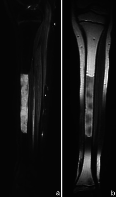
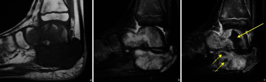
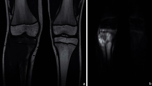
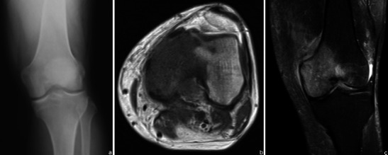
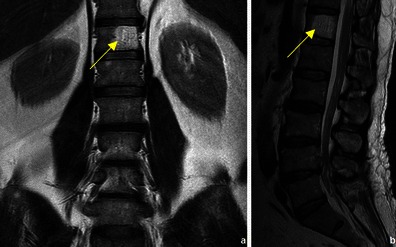
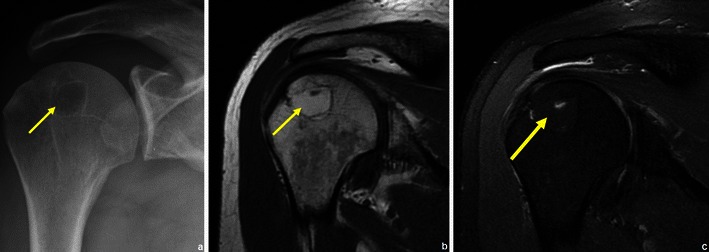
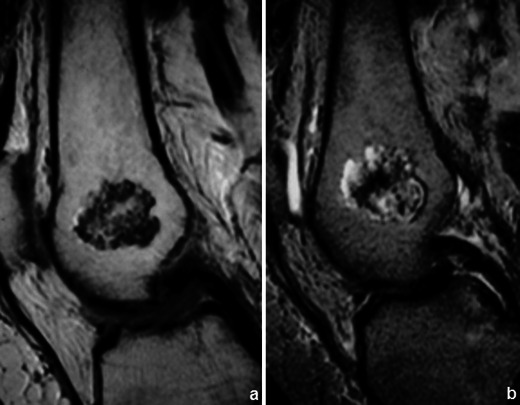
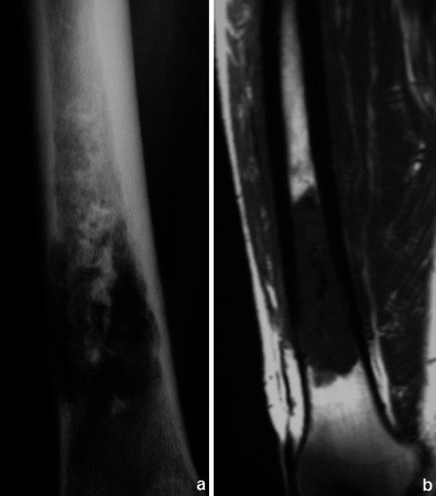
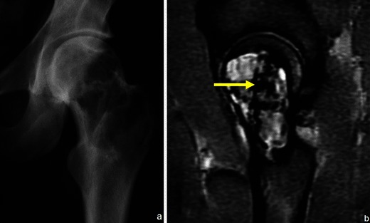
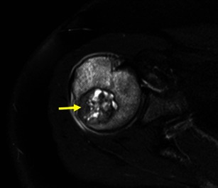
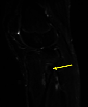
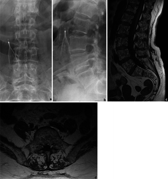
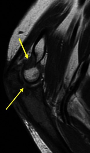
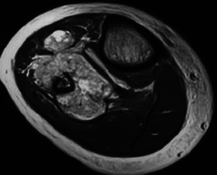
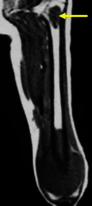
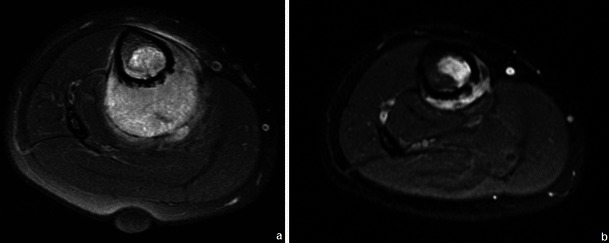
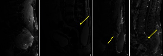
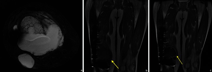
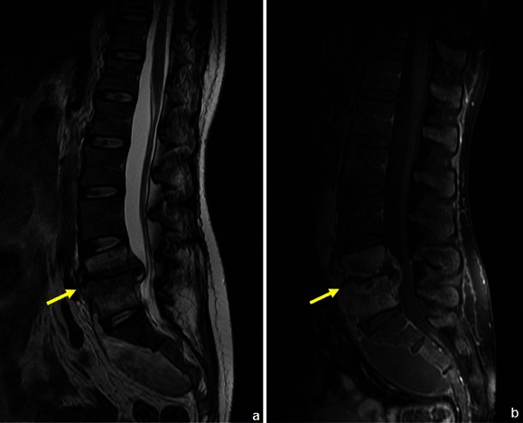
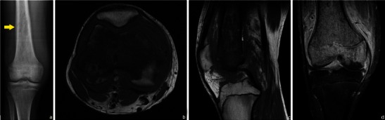
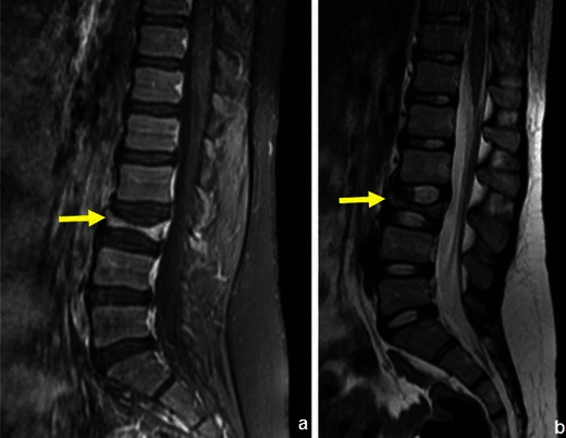
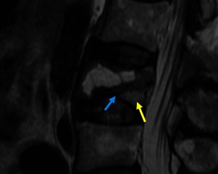
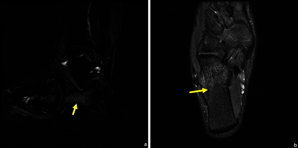
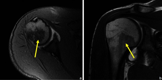
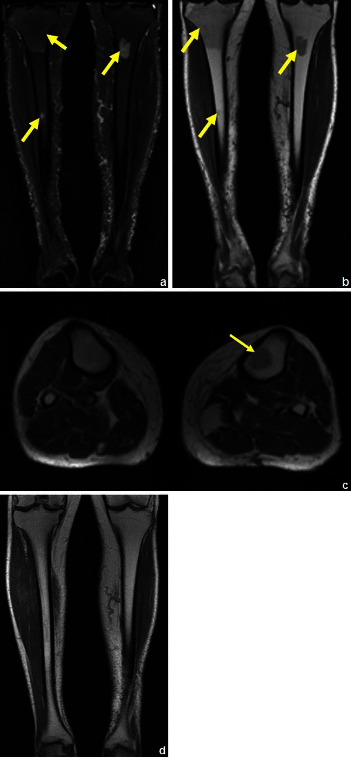
Bone tumours and tumour-like lesions are frequently encountered by radiologists. Although radiographs are the primary screening technique, magnetic resonance imaging (MRI) can help narrow the differential or make a specific diagnosis when a lesion is indeterminate or shows signs of aggressiveness. MRI can extend the diagnostic evaluation by demonstrating several tissue components. Even when a specific diagnosis cannot be made, the differential diagnosis can be narrowed. MRI is superior to the other imaging modalities in detecting bone marrow lesions and tumoral tissue (faint lytic/sclerotic bone lesions can be difficult to visualise using only radiographs). Contrast-enhanced MRI can reveal the most vascularised parts of the tumour and MRI guidance makes it possible to avoid biopsing necrotic areas. MRI is very helpful in local staging and surgical planning by assessing the degree of intramedullary extension and invasion of the adjacent physeal plates, joints, muscle compartments and neurovascular bundles. It can be used in assessing response to neoadjuvant therapy and further restaging. The post-therapeutic follow-up should also be done using MRI. Despite the high quality of MRI, there are a few pitfalls and limitations of which one should be aware. Applications of MRI in bone tumours will probably continue to grow as new sequences are further studied.
• When a lesion is indeterminate or shows signs of aggressiveness, MRI is indicated. • When MRI does not lead to a diagnosis, biopsy is indicated. • MRI is superior to the other imaging modalities in detecting bone marrow lesions. • MRI is very helpful in local staging and surgical planning. • MRI is used in assessing the response to neoadjuvant therapy, restaging and post-therapeutic follow-up.
放射科医生经常会遇到骨肿瘤和肿瘤样病变。虽然 X 线摄影是主要的筛查技术,但当病变不确定或有侵袭性迹象时,磁共振成像(MRI)可以帮助缩小鉴别范围或做出明确诊断。MRI 可以通过显示几种组织成分来扩展诊断评估。即使不能做出明确的诊断,也可以缩小鉴别诊断范围。MRI 在检测骨髓病变和肿瘤组织方面优于其他成像方式(仅使用 X 线摄影很难观察到模糊的溶骨性/硬化性骨病变)。增强 MRI 可以显示肿瘤中最具血管化的部分,并且 MRI 引导可以避免对坏死区域进行活检。MRI 通过评估骨髓内延伸程度以及对相邻骺板、关节、肌肉间隙和神经血管束的侵犯程度,对局部分期和手术计划非常有帮助。它可用于评估新辅助治疗的反应和进一步分期。治疗后的随访也应使用 MRI 进行。尽管 MRI 的质量很高,但还是存在一些需要注意的陷阱和局限性。随着新序列的进一步研究,MRI 在骨肿瘤中的应用可能会继续增加。
当病变不确定或有侵袭性迹象时,应进行 MRI 检查。
如果 MRI 检查不能明确诊断,应进行活检。
MRI 在检测骨髓病变方面优于其他成像方式。
MRI 对局部分期和手术计划非常有帮助。
MRI 用于评估新辅助治疗的反应、重新分期和治疗后的随访。