Zhang Ming, Sun Ze-Qun, Zou Xiao-Ping
Gastroenterology Department, Drum Tower Hospital Affiliated Medical College of Nanjing University, Nanjing, Jiangsu 210008, P.R. China.
Gastroenterology Department, Renmin Hospital, Hubei University of Medicine, Shiyan, Hubei 442008, P.R. China.
Oncol Lett. 2014 Aug;8(2):551-555. doi: 10.3892/ol.2014.2152. Epub 2014 May 19.
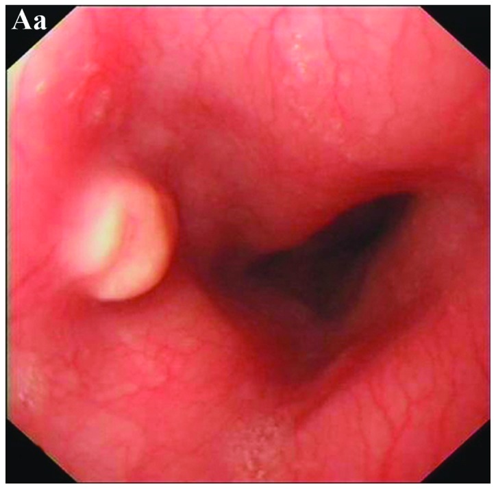
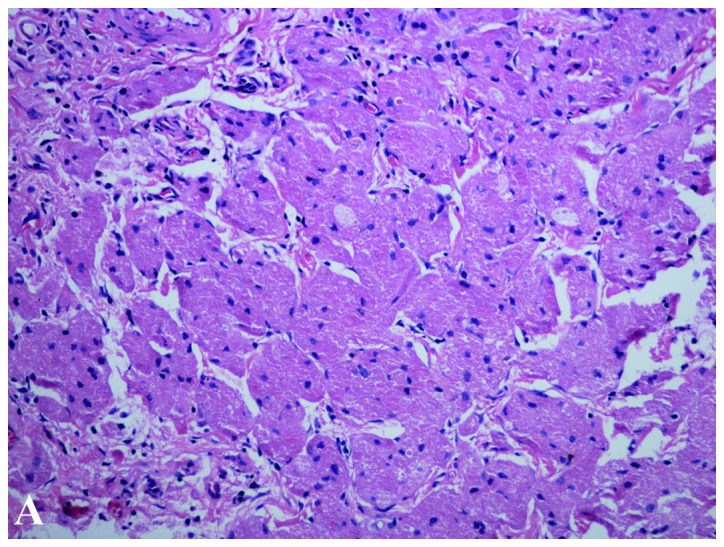
Esophageal granular cell tumors (GCTs) are rare and often misdiagnosed. To demonstrate their clinicopathological features, the present study reports 19 cases and reviews the literature. There were 11 female and eight male esophageal GCT patients with a median age of 42.0 years. All the tumors were solitary. The majority of patient indications for endoscopy (89.5%) were non-specific and endoscopic therapy was performed in 17 cases with a relapse in one case after a 12-month follow-up. The endoscopic appearance of esophageal GCT was variable and the majority of tumors (80.0%) were located in the middle and lower esophageal segments. The size of the tumors ranged from 0.4 to 2 cm in diameter and the surface was white-gray, pink or yellow. Nine patients underwent an endoscopic ultrasound exam, eight of which demonstrated hypoechoic echostructures with a smooth margin and intracavity growth features. One case was derived from the muscularis propria layer with an irregular margin and intra- and extra-cavity growth features. The histological features could mimic other tumors and immunohistochemical stains are usually positive for S-100, periodic acid-Schiff, neuron-specific enolase and nestin. Three cases indicated pleomorphism and Ki-67 was locally positive. Esophageal GCTs are rare and endoscopic ultrasound features are variable. Immunohistochemical staining may aid in the diagnosis.
食管颗粒细胞瘤(GCTs)较为罕见,常被误诊。为阐述其临床病理特征,本研究报告了19例病例并回顾相关文献。19例食管GCT患者中,女性11例,男性8例,中位年龄为42.0岁。所有肿瘤均为单发。大多数患者的内镜检查指征(89.5%)不具有特异性,17例患者接受了内镜治疗,1例在12个月随访后复发。食管GCT的内镜表现各异,大多数肿瘤(80.0%)位于食管中下段。肿瘤直径为0.4至2厘米,表面呈灰白色、粉红色或黄色。9例患者接受了内镜超声检查,其中8例显示为低回声结构,边界光滑,具有腔内生长特征。1例起源于固有肌层,边界不规则,具有腔内和腔外生长特征。其组织学特征可类似其他肿瘤,免疫组化染色通常S-100、过碘酸希夫反应、神经元特异性烯醇化酶和巢蛋白呈阳性。3例显示多形性,Ki-67局部呈阳性。食管GCT较为罕见,内镜超声特征各异。免疫组化染色可能有助于诊断。