Neuroscience Laboratory, Departments of Neurology and Pediatrics, Hugo Moser Research Institute at Kennedy Krieger, Johns Hopkins University School of Medicine Baltimore, MD, USA.
Neuroscience Laboratory, Hugo Moser Research Institute at Kennedy Krieger Baltimore, MD, USA.
Front Syst Neurosci. 2014 Jun 27;8:118. doi: 10.3389/fnsys.2014.00118. eCollection 2014.
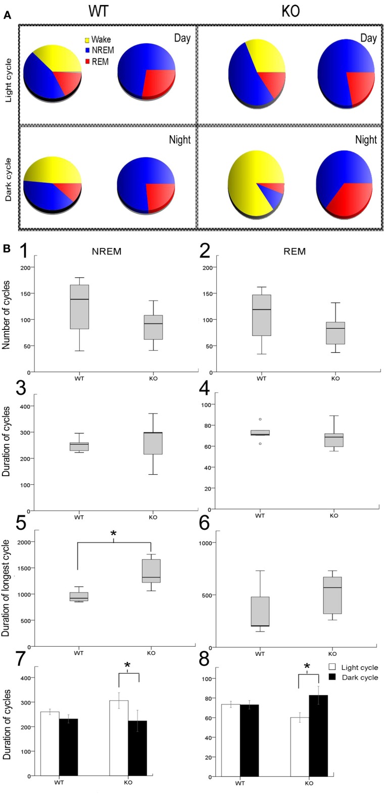
Mutations in the X-linked gene encoding methyl-CpG-binding protein 2 (Mecp2) cause most cases of Rett syndrome (RTT). Currently there is no cure for RTT. Abnormal EEGs are found in 100% of RTT cases and are associated with severe sleep dysfunction, the cause of which is not well understood. Mice deficient in MeCP2 protein have been studied and characterized for their neuropathological and behavioral deficits to better understand RTT. With the goal to study the non-ictal EEG correlates in symptomatic Mecp2 KO mice (Mecp2(tm1.1Bird/y)), and determine novel EEG biomarkers of their reported progressive neurodegeneration, we used 24 h video-EEG/EMG with synchronous in-vivo cortical glutamate biosensor in the frontal cortex. We scored the EEG for activity states and spectral analysis was performed to evaluate correlations to the synchronous extracellular glutamate fluctuations underlying Mecp2 inactivation as compared to WT. Significant alterations in sleep structure due to dark cycle-specific long wake states and poor quality of slow-wave sleep were associated with a significant increase in glutamate loads per activity cycle. The dynamics of the activity-state-dependent physiological rise and fall of glutamate indicative of glutamate homeostasis were significantly altered in the KO mice. Colorimetric quantitation of absolute glutamate levels in frontal cortex also indicated the presence of significantly higher levels in KO. This study for the first time found evidence of uncompensated sleep deprivation-like EEG biomarkers that were associated with glutamate homeostatic dysfunction in the Mecp2 KO mice.
X 连锁基因编码甲基-CpG 结合蛋白 2(Mecp2)的突变导致大多数雷特综合征(RTT)病例。目前,RTT 尚无治愈方法。100%的 RTT 病例中发现异常脑电图,与严重的睡眠功能障碍有关,但原因尚不清楚。已经对缺乏 MeCP2 蛋白的小鼠进行了研究,并对其神经病理学和行为缺陷进行了特征描述,以便更好地了解 RTT。为了研究有症状的 Mecp2 KO 小鼠(Mecp2(tm1.1Bird/y))中的非癫痫发作性 EEG 相关性,并确定其报道的进行性神经退行性变的新 EEG 生物标志物,我们使用了 24 小时视频-EEG/EMG 与同步活体皮质谷氨酸生物传感器在前额叶皮层。我们对 EEG 进行了活动状态评分,并进行了频谱分析,以评估与同步细胞外谷氨酸波动的相关性,这种波动是由于 Mecp2 失活而导致的,与 WT 相比。由于暗周期特定的长时间清醒状态和慢波睡眠质量差,导致睡眠结构发生显著改变,与每个活动周期的谷氨酸负荷显著增加有关。表明谷氨酸稳态的活动状态依赖性生理上升和下降的动力学在 KO 小鼠中发生了显著改变。前皮层绝对谷氨酸水平的比色定量也表明 KO 中的谷氨酸水平明显升高。这项研究首次发现了证据表明存在未补偿的睡眠剥夺样 EEG 生物标志物,与 Mecp2 KO 小鼠中的谷氨酸稳态功能障碍有关。