Wüstner Daniel, Christensen Tanja, Solanko Lukasz M, Sage Daniel
Department of Biochemistry and Molecular Biology, University of Southern Denmark, DK-5230 Odense M, Denmark.
Biomedical Imaging Group, Ecole Polytechnique Fédérale de Lausanne (EPFL), CH-1015 Lausanne, Switzerland.
Molecules. 2014 Jul 29;19(8):11096-130. doi: 10.3390/molecules190811096.
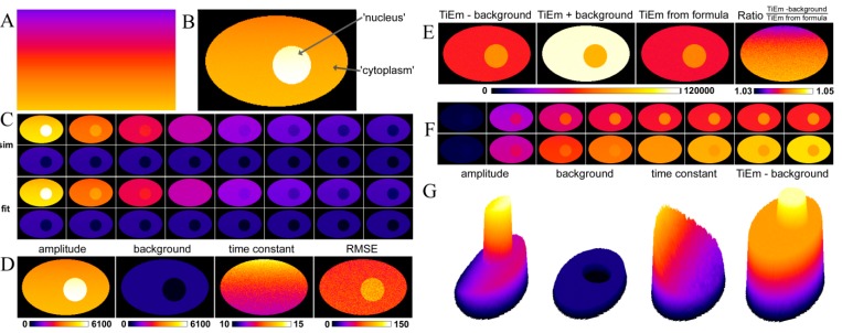
Since the pioneering work of Hirschfeld, it is known that time-integrated emission (TiEm) of a fluorophore is independent of fluorescence quantum yield and illumination intensity. Practical implementation of this important result for determining exact probe distribution in living cells is often hampered by the presence of autofluorescence. Using kinetic modelling of photobleaching combined with pixel-wise bleach rate fitting of decay models with an updated plugin to the ImageJ program, it is shown that the TiEm of a fluorophore in living cells can be determined exactly from the product of bleaching amplitude and time constant. This applies to mono-exponential bleaching from the first excited singlet and/or triplet state and to multi-exponential combinations of such processes. The TiEm can be used to correct for illumination shading and background autofluorescence without the need for fluorescent test layers or separate imaging of non-stained cells. We apply the method to simulated images and to images of cells, whose membranes were labelled with fluorescent sterols and sphingolipids. Our bleaching model can be extended to include a probability density function (PDF) of intrinsic bleach rate constants with a memory kernel. This approach results in a time-dependent bleach rate coefficient and is exemplified for fluorescent sterols in restricted intracellular environments, like lipid droplets. We show that for small deviations from the classical exponential bleaching, the TiEm of decay functions with rate coefficients remains largely independent of fluorescence lifetime and illumination, and thereby represents a faithful measure of probe distribution.
自赫希菲尔德的开创性工作以来,人们就知道荧光团的时间积分发射(TiEm)与荧光量子产率和光照强度无关。在活细胞中确定精确的探针分布时,这一重要结果的实际应用常常受到自发荧光的阻碍。通过将光漂白的动力学模型与使用ImageJ程序的更新插件对衰减模型进行逐像素漂白速率拟合相结合,结果表明,活细胞中荧光团的TiEm可以从漂白幅度和时间常数的乘积精确确定。这适用于从第一激发单重态和/或三重态的单指数漂白以及此类过程的多指数组合。TiEm可用于校正光照阴影和背景自发荧光,而无需荧光测试层或对未染色细胞进行单独成像。我们将该方法应用于模拟图像和细胞膜用荧光固醇和鞘脂标记的细胞图像。我们的漂白模型可以扩展到包括具有记忆核的固有漂白速率常数的概率密度函数(PDF)。这种方法产生一个随时间变化的漂白速率系数,并以受限细胞内环境(如脂滴)中的荧光固醇为例进行说明。我们表明,对于与经典指数漂白的小偏差,具有速率系数的衰减函数的TiEm在很大程度上仍与荧光寿命和光照无关,因此是探针分布的可靠度量。